THE RUMEN AND RETICULUM
The rumen and reticulum together form the vessel in which the unpromising food material, invulnerable to attack by mammalian digestive enzymes, is reduced by processes of microbial fermentation.
Some of the simpler products are assimilated directly, while others are susceptible to conventional digestion lower in the digestive tract.The rumen is laterally compressed and extends from the cardia—which lies a little way above the middle of the seventh intercostal space or eighth rib—to the pelvic inlet, from the abdominal roof to the floor, and from the left body wall across the midline, especially caudally and ventrally, where it may reach the lower right flank (see Figure 28-12). The much smaller reticulum lies cranial to the rumen under cover of the sixth to eighth ribs and mainly to the left of the median plane. It reaches from the cardia to the most forward part of the diaphragm and occupies the full height of this shallower part of the abdomen; it also passes across the midline, especially ventrally, where it lies above the xiphoid process of the sternum (see Figures 27-7/8 and 28-4/5).
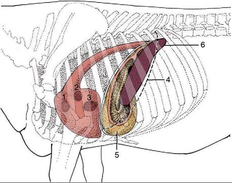
Figure 28-5 Left lateral projection of certain organs on the bovine thoracic wall. 1, Pulmonary valve; 2, aortic valve; 3, left atrioventricular valve; 4, position of basal border of the lung; 5, reticulum, opened (note position of reticular groove); 6, spleen.
This position allows the application of external pressure in the expectation of eliciting pain when the reticulum is diseased.
The rumen and reticulum are so intimately related in structure and function that many now prefer to describe a combined ruminoreticular compartment. There is much in favor of this convention. The division of the rumen from the reticulum, though more complete, is
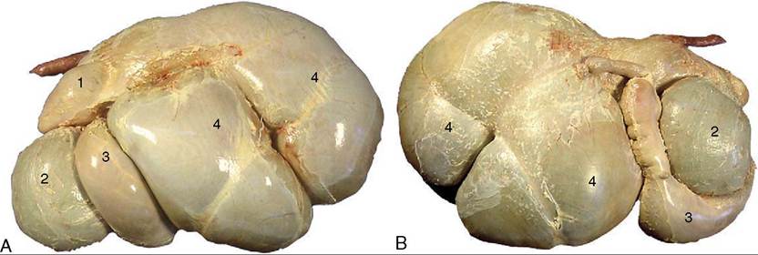
Figure 28-7 A, Bovine stomach, left side.
B, Bovine stomach, right side. 1, Reticulum; 2, omasum; 3, abomasum; 4, rumen.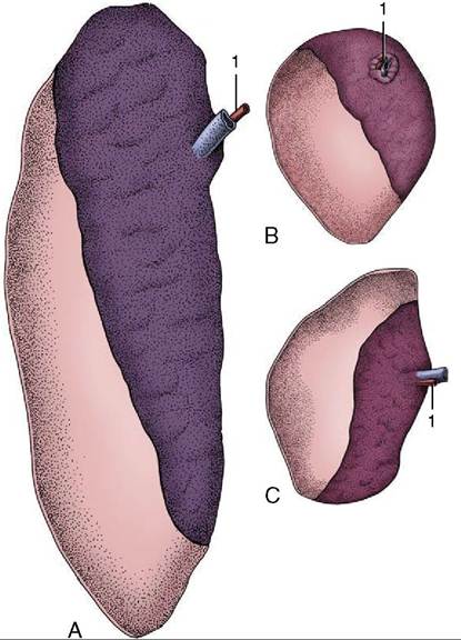
Figure 28-6 The spleens of cattle (A), sheep (B), and goats (C), visceral surface. The craniodorsal area is bare. The splenic artery (1) is indicated.
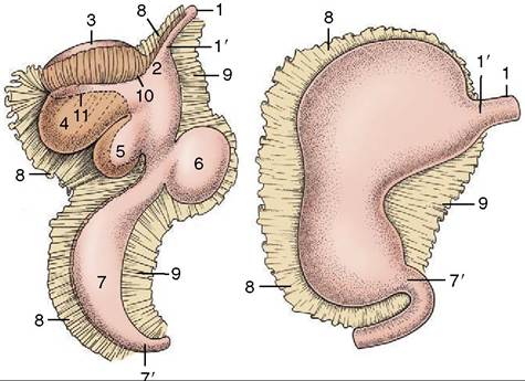
Figure 28-8 The attachments of the greater and lesser omenta on the developing ruminant stomach. The simple stomach to the right shows the correspondence of its parts to the compartments of the ruminant stomach. 1, Esophagus; 1', cardia; 2, atrium ruminis; 3, dorsal sac of rumen; 4, ventral sac of rumen; 5, reticulum; 6, omasum; 7, abomasum; 7’, pylorus; 8, greater omentum; 9, lesser omentum; 10, part of greater curvature corresponding to the right longitudinal groove of the rumen; 11, part of greater curvature corresponding to the left longitudinal groove of the rumen.
achieved in exactly the same way as the subdivision of the rumen, namely by the inflection of the walls to form a series of pillars (pilae) that project internally. The whole thickness of the stomach wall, except the peritoneum, participates in these formations. The divisions and the pillars that bound them are illustrated in Figure 28-4, B. The rumen and reticulum communicate over the U-shaped ruminoreticularfold. The principal ruminal pillars encircle the organ, dividing dorsal and ventral major sacs, while lesser coronary pillars mark off the caudal blind sacs. The cranial pillar has an oblique direction that partially divides the cranial extremity from the remainder of the dorsal sac, emphasizing the association of the former part (atrium ruminis) with the reticulum. External grooves correspond to the positions of all these folds. The relative proportions of the compartments vary among the domestic ruminants. The smaller size of the dorsal sac and the extensive caudal projection of the ventral blind sac give the rumen of sheep and goats an unbalanced appearance when compared with the more symmetrical bovine rumen.
There are also differences in the development of the grooves visible externally, but these are altogether without significance.The serosa covers the entire surface of the rumen and reticulum, except dorsally where the ruminal wall is directly adherent to the abdominal roof from the esoph-
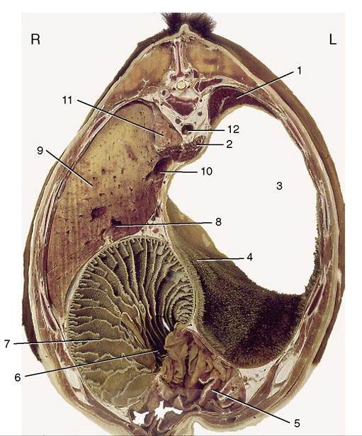
Figure 28-9 Transverse section of the bovine trunk at the level of the tenth thoracic vertebra. 1, Spleen; 2, crura of diaphragm; 3, atrium ruminis; 4, cranial pillar; 5, abomasum; 6, omasoabomasal opening; 7, omasum; 8, portal vein; 9, liver; 10, caudal vena cava; 11, right lung; 12, aorta.
ageal hiatus of the diaphragm to the level of the fourth lumbar vertebra (Figure 28-13/22), and over certain grooves where it is reflected to continue into the greater omentum. The limited attachment allows the ruminore- ticulum the freedom necessary for the incessant and reciprocal contractions and enlargements of its various parts.
The relationships are most easily studied by reference to the illustrations (see Figures 28-4, A-B; 28-7; and 28-10). The most important points are contact between the reticulum and the diaphragm and liver cranially; insinuation of the abomasum between the two chambers (ventral sac of rumen and reticulum) ventrally; relation of the right surface of the rumen to the intestinal mass, omasum, abomasum, pancreas, and kidneys; and the intrusion of the superficial wall of the greater omentum between the ventral sac of the rumen and the abdominal wall. The rumen also has a variable relationship to the uterus and other organs at the entrance to the pelvis, where the dorsal sac may be palpated per rectum. The direct contact of the dorsal sac with the upper part of the left flank makes auscultation and
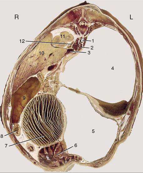
Figure 28-10 Transverse section of the bovine trunk at the level of the thirteenth thoracic vertebra.
1, Aorta; 2, right crus of diaphragm; 3, caudal vena cava; 4, dorsal sac of rumen; 5, ventral sac of rumen; 6, abomasum; 7, omasum; 8, duodenum; 9, gallbladder; 10, liver; 11, cranial pole of right kidney; 12, right adrenal gland.palpation simple. It also facilitates trocarization for the relief of tympany.
The interior of the ruminoreticulum communicates with the esophagus and omasum through openings placed at the extremities of the reticular groove, a prominent gutter that descends from the cardia over the right face of the reticulum toward the fundus (Figure 28-14Z√,5). The groove is bounded by spiral fleshy lips; the upper end of the left (cranial) lip is expanded to overhang the slitlike cardiac opening, while a similar thickening of the lower end of the right (caudal) lip partly conceals the round exit into the omasum. The cardia is placed at the junction of the rumen and reticulum and discharges into both chambers. In the unweaned animal the reticular groove may be converted into a closed tube, forming a channel that conveys milk directly from the esophagus to the omasal canal, whence it drops into the abomasum. The muscular contractions that draw the lips together are reflexly stimulated by sucking from the dam or by the presentation of suitable bucket feeds. As the animal matures, alterations in diet and feeding regimen result in decreasing use of this route, although even in the adult a portion of the soluble nutrients released into the saliva during mastication
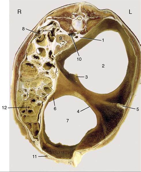
Figure 28-11 Transverse section of the bovine trunk at the level of the third lumber vertebra. 1, Aorta: 2, caudodorsal blind sac; 3, dorsal coronary pillar; 4, caudal pillar; 5, left longitudinal pillar; 6, ventral coronary pillar; 7, caudoventral blind sac; 8, descending duodenum; 9, left kidney; 10, caudal vena cava; 11, milk vein; 12, intestinal mass.
succeeds in bypassing the ruminoreticulum.
The groove reflex is stimulated by antidiuretic hormone (ADH), which indicates that the reflex may have some function in adult life. ADH is produced in response to dehydration or an increase in plasma osmolality. ADH is associated with thirst, and its effect on the reticular groove may cause a portion of the water drunk by dehydrated animals to bypass the ruminoreticulum. Closure of the groove can be stimulated by certain chemicals (e.g., copper sulfate). This provides a useful strategy when it is desirable to introduce drugs to the abomasum without prior dilution in the forechambers.The ruminoreticular mucosa is lined by a harsh stratified cutaneous epithelium (Figure 28-15, A-B) that is stained a greenish brown; the floor of the reticular groove, however, is smooth and pale. The reticular mucosa has a distinctive pattern formed by ridges about 1 cm high that outline four-, five-, and six-sided “cells” (see Figures 27-7Z8 and 28-16, B). These ridges and the cell floors between them carry low papillae. The reticulate pattern becomes less regular toward the junction with the rumen and gradually modifies to merge with
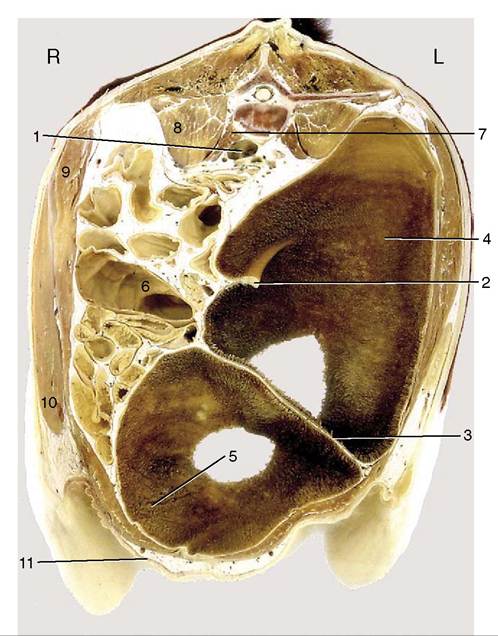
Figure 28-12 Transverse section of the bovine trunk at the level of the fifth lumbar vertebra. 1, Bifurcation of aorta and formation of caudal vena cava; 2, right dorsal coronary pillar; 3, caudal pillar; 4, caudodorsal blind sac; 5, caudoventral blind sac; 6, colon; 7, psoas minor; 8, psoas major; 9, internal abdominal oblique; 10, external abdominal oblique; 11, milk vein.
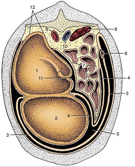
Figure 28-13 Schematic transverse section of the abdominal cavity to show the disposition of the greater omentum. 1, Dorsal sac of rumen; 2, ventral sac of rumen; 3, superficial wall of greater omentum; 4, deep wall of greater omentum; 5, omental bursa; 6, descending duodenum; 7, intestinal mass; 8, right kidney; 9, aorta; 10, caudal vena cava; 11, supra- omental recess; 12, retroperitoneal attachment of rumen.
the papillated surface of this chamber. The upper keratinized layer of the epithelium protects against abrasion by the rough, fibrous diet, whereas the deeper layers metabolize volatile short-chain fatty acids. Histologically, the epithelium shows many similarities with the epidermis. The lamina propria-submucosa, formed by a network of collagen and elastic fibers, includes bands of smooth muscle within the distal parts of the reticular ridges (Figure 28-15, A). The ruminal papillae vary in prominence according to age, diet, and location (Figure 28-16, A, and Figure 28-14). Normally they are largest and most densely strewn within the blind sacs, fewer and less prominent in the ventral sac, and least developed over the center of the roof and toward the free margins of the pillars. Individual papillae vary from low rounded elevations through conical and tonguelike forms to flattened leaves about 1 cm long. The ruminal epithelium resembles that of the reticulum. A thick lamina propria beneath the epithelium forms the core of the papilla; apart from collagen, elastic, and reticular fibers, it includes a dense capillary network. There is no muscularis mucosae. The looser submucosa is located directly against the lamina propria and also contains a vascular network (Figure 28-15, B).
The rugose nature of the ruminoreticular lining was formerly interpreted as an adaptation for the mechanical disruption of the macerating ingesta. Since it became known that the volatile fatty acids produced by microbial fermentation are absorbed in the rumen and reticulum, it has been regarded as primarily a device to increase the epithelial surface. Papillary development is stimulated by these acids (especially butyric), and their absorption is facilitated by the very rich subepithelial capillary plexus. In some wild ruminants, striking changes in papillary prominence and size, and thus in the ruminal surface area (Figure 28-16, A), accompany seasonal changes in forage quality. Changes in papillary development tend to be more restrained in domestic species, whose diet is subject to human influence to a greater or lesser degree.
The reticulum of the small ruminants is relatively larger than that of cattle. Although it extends farther caudally, its contact with the abdominal floor is subject to much functional variation (Figure 28-14/5). There are conspicuous species differences in its lining. The ridges that bound the reticular “cells” are relatively much lower and have more prominently serrated
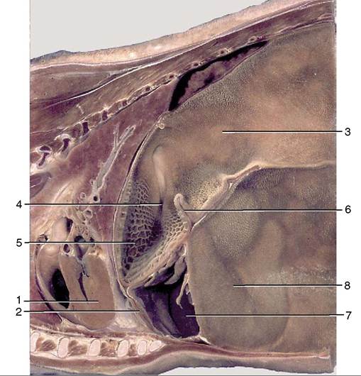
Figure 28-14 Paramedian section of part of the trunk of a goat. 1, Heart; 2, diaphragm; 3, atrium ruminis; 4, reticular groove; 5, reticulum; 6, ruminoreticular fold; 7, abomasum; 8, ventral sac of rumen.
margins. The papillated “ruminal” mucosa also extends over a larger part of the reticular wall.
The smooth muscle of the ruminoreticular wall is arranged in two coats that continue the striated muscle of the esophagus. The thin outer coat runs craniocau- dally over the rumen but has an oblique course on the reticulum. Most bundles of the much thicker inner layer run more or less at right angles to the superficial coat and thus encircle the long axis of the rumen. They extend into the pillars and form the bases of these structures. The thicker parts of the ruminoreticular muscle are sold for consumption as tripe.
The regular sequence of ruminoreticular contractions mixes and redistributes the stomach contents. The cycle consists of a biphasic reticular contraction (relaxation between contraction phases is more consistent in cattle than in sheep), which throws the reticular contents into the atrium ruminis, followed by contraction of first the dorsal and later the ventral rumen sacs. The wave of contraction passes over each in a craniocaudal direction. The process is centrally regulated, and the tempo and vigor are adjusted according to information supplied by intramural receptors that are stimulated by stretching of the wall and by contact with floating fragments. Both the sensory and the motor pathways travel within the vagus nerves.
Regurgitation of food for remastication requires the coordination of the stomach movements with those of the thoracic wall and throat. It is preceded by an additional reticular contraction that floods the cardiac region; the ingesta are drawn into the esophagus on expansion of the thorax with a closed upper airway and are then carried orally by an antiperistaltic wave. The heavy remasticated cud, now further sodden and divided, tends to drop from the cardia into the reticulum.
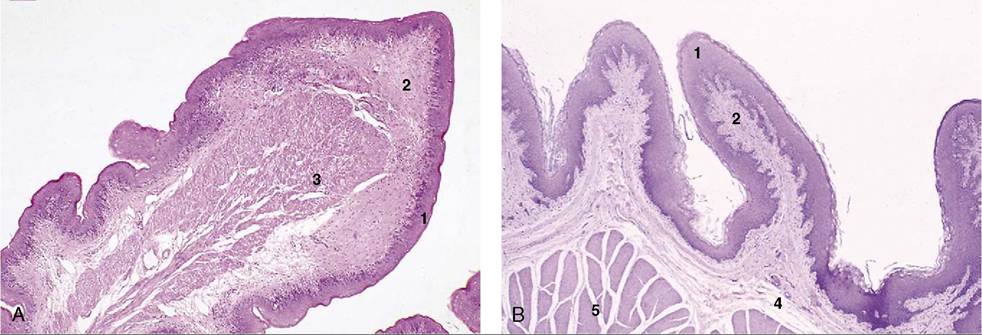
Figure 28-15 A, Reticulum (goat) (28?). B, Rumen (goat) (28?). 1, Stratified squamous epithelium; 2, lamina propria; 3, lamina muscularis mucosae; 4, submucosa; 5, muscularis interna.
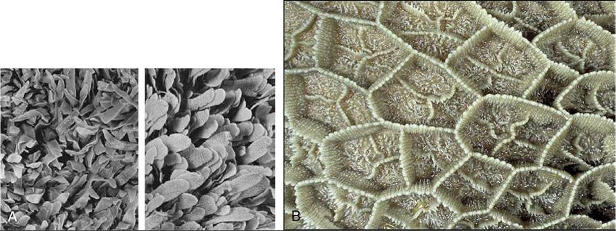
Figure 28-16 A, Rumen papillated mucosa taken from a Waterbuck (left) and a lesser Kudu. B, Reticulum: mucosal ridges outlining "cells" characteristic of the reticular mucosa (cow).
In eructation (the discharge of gas through the esophagus), ruminal contractions in which the reticulum does not participate are substituted for the normal pattern of activity. These contractions originate in the ventral sac and generally spread to the dorsal sac, where they begin caudally and extend cranially; they force the ruminal gas forward to the cardiac area whence it is aspirated into the esophagus, through which it is hurried orally by an antiperistaltic wave. It then passes through the relaxed pharyngoesophageal sphincter into the pharynx. Some escapes from the mouth, but part is directed to the lungs.
The content of the rumen shows some stratification: food of recent ingestion is piled above the heavier, more sodden remasticated material. It is therefore the lighter material that is most liable to be regurgitated for further mastication and insalivation (Figure 28-17).
Cattle are notoriously careless feeders and often ingest foreign bodies, especially pieces of wire, with their forage. These bodies tend to collect within the reticulum and, when sharp, may be driven through the reticular wall by the contractions of this organ (traumatic reticulitis—“hardware disease”). Common sequelae include abscessation of the liver and possibly other abdominal organs and, more critically, a purulent pericarditis when the object penetrates the diaphragm. Some of these bodies corrode, while others may be immobilized by introducing a magnet through the mouth (Figure 28-18/2 and inset).