THE SIMPLE STOMACH
1. What is the principal tissue of the esophagus? Describe its principal features (consider accommodation of a large bolus or other object). On which side of the neck could a bolus in the esophagus be observed?
2.
What are the glandular regions of the stomach, and what are their secretions?3. Note the extent of the glandular and nonglandular regions of the stomach (see Fig. 12-2).
4. What are the forestomachs of the ruminant? What is their function?
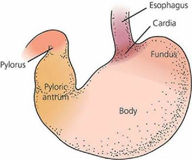
■ FIGURE 12-6 Parts of the canine stomach.
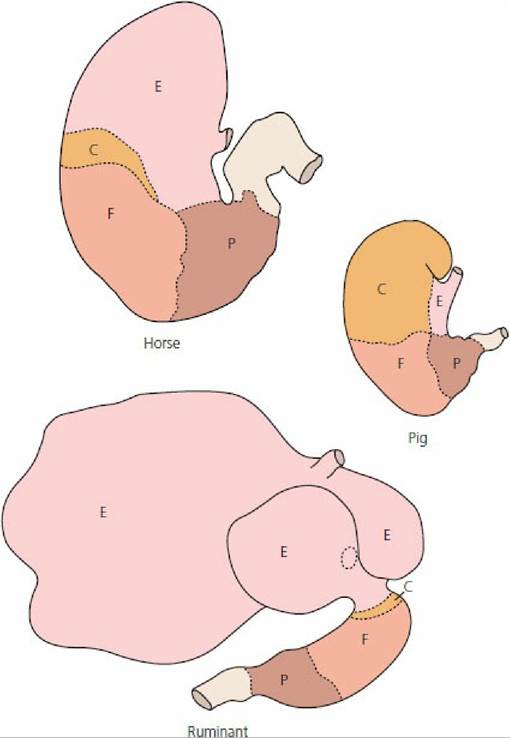
■ FIGURE 12-7 Inner regions of the stomach in the horse, pig, and ruminant. E, Nonglandular region; C, cardiac gland region; F, fundic gland region; P, pyloric gland region. (From Frandson RD, Wilke WL, Fails AD. Anatomy and Physiology of Farm Animals. 7th edn. Ames IA: Wiley-Blackwell, 2009.)
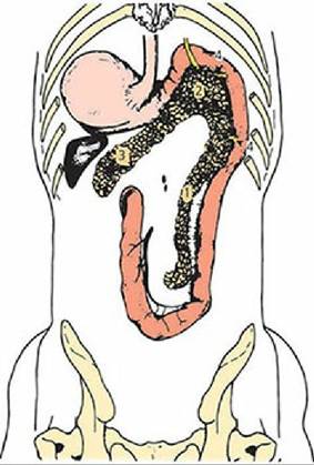
■ FIGURE 12-8 Dorsal view of the canine stomach, duodenum, and pancreas. 1, Right lobe of pancreas; 2, body of the pancreas; 3, left lobe of pancreas; 4, pancreatic ducts. The common bile duct is received into the duodenum in close association with the pancreatic duct. (From Adams DR. Canine Anatomy: A Systemic Approach. Ames, IA: Iowa State University Press, 1986.) Regarding stomach structure and function, domestic animals are represented by two general classes, ruminants and nonruminants. Cattle, sheep, and goats belong to the former class; the other domestic animals belong to the latter. The stomach of nonruminants is relatively simple, consisting of only one compartment. For this reason it is frequently referred to as the simple stomach.
The stomach of birds is unique and will be described separately. That of ruminants is more complex, consisting typically of four compartments, only one of which secretes gastric juice. Digestion in the simple stomach will be described first, with some references to ruminants, but the major aspects of ruminant digestion are described separately.Esophagus
The esophagus is a muscular tube extending from the pharynx to the stomach. During its course to the stomach, the esophagus enters the thorax at the thoracic inlet and travels within the mediastinal space, in which it is subjected to pressure changes associated with that space. The esophagus finally passes through its opening in the diaphragm and enters the stomach within the abdominal cavity. Food and water are moved from the pharynx to the stomach by contraction waves in the muscular wall. Esophageal sphincters (points at which flow is controlled) have not been morphologically shown, but have been suggested by functional studies Muscle activity can constrict the lumen of the esophagus at certain locations. Accordingly, the esophagus is normally closed at the pharyngeal end by tonic muscle activity providing for a cranioesophageal sphincter. Although a thickening, suggestive of a sphincter, occurs at the junction of the esophagus with the stomach (the cardia), the opening remains closed, not because of an anatomic sphincter but because of a closure that is functional in nature. The lumen of the esophagus is normally closed, which produces folds in its inner surface. During passage of a bolus the folds are extended so that a minimum of stretch is necessary. Unusually large objects extend the folds and stretch the mucosal and submucosal layers, and they can become lodged at points of narrowing (e.g., the thoracic inlet).
The pharyngeal opening to the esophagus lies just above the glottis, which is the opening to the trachea. On its way to,the stomach the esophagus courses along the left side of the trachea in the neck region.
Bolus transport can be observed by watching the left side of the neck (this is particularly apparent in cattle).The muscle fibers of the esophagus are arranged circularly and longitudinally. In most animals they are striated, but a part of the caudal portion is smooth muscle in some. Because of the muscular nature of the esophagus, it is recovered at slaughter as an edible source for certain meat products.
Stomach
Food is received by the stomach for storage (pending further digestion) and for the beginning of digestion. Because the stomach serves a storage function, it is a dilated portion of the digestive tube. As viewed from the outside (Figure 12-6), it is seen to be subdivided into parts, which are continuous with one another. The cardia (entrance area) is located nearest the esophagus and is continued by the fundus, which is the dome-shaped part of the stomach. The fundus is adjacent to the body, which is the large middle portion. It extends from the fundus to the pyloric antrum. The pyloric antrum is the constricted part of the stomach that joins with the duodenum via the pylorus (a sphincter muscle that controls stomach emptying). The lesser curvature is the very short side of the stomach between the cardia and pylorus. The greater curvature defines the much longer convex opposite side.
Depending on the species, the interior of the stomach can be subdivided into glandular and nonglandular (esophageal) regions. The carnivore is an example of an animal that has a stomach that is entirely glandular, while that of the horse has both glandular and nonglandular areas. The glandular part of the stomach has specific regions according to cell type: cardiac gland, fundic gland, and pyloric gland regions. The several regions of the stomach lining for the horse, pig, and ruminant are shown in Figure 12-7. The fundic gland region includes the entire space between the cardiac gland (nearest the cardia) and pyloric gland regions (near the pylorus); these glands are sometimes called the gastric glands.
The cardiac, gastric, and pyloric glands all secrete mucus. In addition, the gastric glands secrete hydrochloric acid (HCl) and pepsinogen by their parietal and neck chief cells, respectively. The pyloric glands also secrete the hormone gastrin.The nonglandular region of the stomach is covered by stratified squamous epithelium and has no secretory capacity. It varies in size, depending on the species, and can be confined to the area immediately around the esophageal opening or make up a larger percentage of the stomach. Notice the differences for the ruminant, whereby the ruminant forestomachs (the rumen, reticulum, and omasum) are followed by the true stomach, or abomasum. The forestomachs comprise the entire nonglandular region, and the epithelium of the abomasum consists mostly of the fundic gland region and pyloric gland region (glandular part of the stomach). The cardiac gland region occupies a very small area adjacent to the opening into the abomasum from the omasum.
■