INTESTINES
1. What part of the small intestine receives the pancreatic and bile duct?
2. For those animals not requiring extensive fermentation of their food, where does most of the digestion and absorption take place?
3.
Study Figures 12-9,12-10,12-11,12-12, and 12-13. These explain and illustrate the functional aspects of small intestine morphology. Read the text that accompanies these figures.4. How is the surface of the small intestine amplified?
5. How are epithelial cells for the.villi renewed? What is their replacement time?
6. How does blood and lymph return from the intestines differ?
7. Is fermentation common to the large intestine of all animals? What is the location difference for fermentation between ruminant and nonruminant herbivores?
8. Are the microbes of fermentation available for their own digestion in both ruminant and nonruminant herbivores?
9. Note the differences of the digestive tract between the cecum and transverse colon among the domestic animals. Which animals have an ansa spiralis? Which animal has the double horseshoe (ventral and dorsal large colon)?
0. What is the function of the sacculations (haustra) in the cecum and colon of the pig and horse?
11. What is the rectum?
268" class="lazyload" data-src="/files/uch_group31/uch_pgroup304/uch_uch7236/image/image265.jpg">
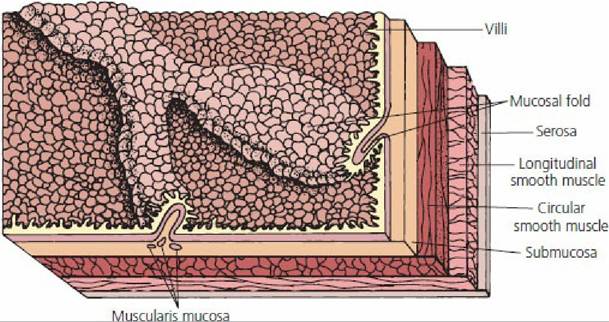
■ FIGURE 12-9 Schematic representation of the general organizational features of the mammalian gastrointestinal tract. A. Cross-section of the small intestine with its mesenteric suspension that envelops the intestine as its serosa. B. Boxed section from A to show greater detail. Auerbach’s nerve plexus controls gastrointestinal movements. Meissner’s plexus (not shown) is in the submucosa, and controls secretions and blood flow. The muscularis mucosae produces folds in the mucosa for amplification of the surface area.
■ FIGURE 12-10 A layered section of intestine as viewed from its inner surface.
The folds are produced by strategic contraction of the muscularis mucosae. The projections from the surface represent the villi, another means of surface amplification.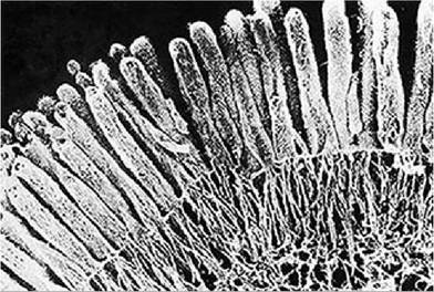
■ FIGURE 12-11 Photomicrograph of microvilli extending from a small intestine epithelial cell. The cord-like structures extending downward from the microvilli are contractile actin filaments. (From Fawcett DW. Bloom and Fawcett: A Textbook of Histology. 11th edn. Philadelphia, PA: W.B. Saunders, 1986. Courtesy of N. Hirokawa and J. Heuser.)
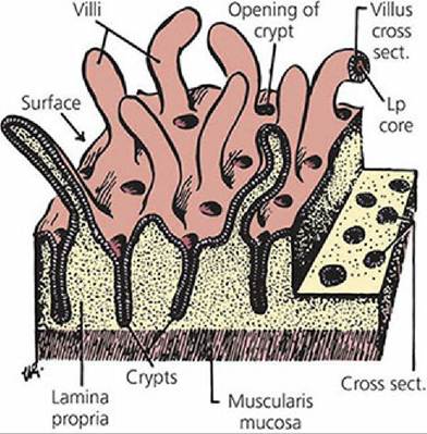
■ FIGURE 12-12 Three-dimensional representation of the small intestine lining. The villi are finger-like processes with cores of lamina propria that extend into the lumen. The crypts of Lieberkuhn,are depressions into the lamina propria (Lp). (From Ham AW. Histology. 7th edn. Philadelphia, PA: J.B..Lippincott, 1974.)
B
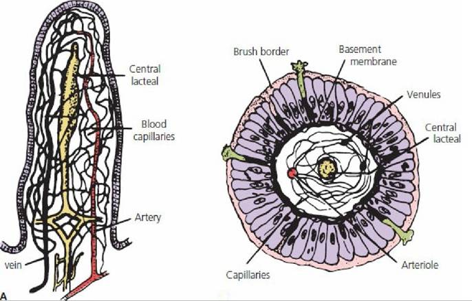
■ FIGURE 12-13 Functional organization of the villus: A. Longitudinal section. B. Cross-section showing the epithelial cells and basement membrane. (From Guyton AC. Textbook of Medical Physiology. 8th edn. Philadelphia, PA: WB Saunders, 1991.)
Content leaving the stomach and entering the intestine is known as chyme. Its consistency is fluid or semifluid and its reaction is acid. The composition of chyme depends on the diet and feeding habits of the animal. In the intestine, chyme undergoes important changes, which constitute intestinal digestion.
Small Intestine
The small intestine is composed of three sections as it proceeds caudally from the pylorus: the duodenum, jejunum, and ileum. The duodenum makes a loop as it turns to cross from the right to the left side. Closely related to the duodenum is the pancreas. The duodenum receives pancreatic secretions involved in digestion through two pancreatic ducts for most species and one for others (Figure 12-8).
The duodenum also receives bile formed in the liver through the common bile duct, which transports bile from the liver or gallbladder to the intestine. Most digestion and absorption takes place in the small intestine for those animals not requiring extensive fermentation of their ingested food. A cross-section of the small intestine is shown in Figure 12-9.The inner layer of the small intestine, having intimate contact with the contents of the lumen, is composed of an epithelial cell layer known as the mucosa. The submucosa is a connective tissue layer that provides space for blood vessels, lymph vessels, and nerve fibers. In addition, a sparse layer of smooth muscle fibers is in the submucosa, known as the muscularis mucosae. The muscularis mucosae produce folds in the mucosa, thereby increasing surface area. These folds change location to bring different parts of the intestine into more intimate contact with the luminal contents. Individual fibers from the muscularis mucosae attach to villi and cause villus movement when contracting. This facilitates lymph movement and placement of the villus into new areas of luminal fluid. Beneath the submucosa are circular and longitudinal muscle layers composed of smooth muscle fibers. Contraction of these muscles is associated with mixing and propulsive movements of intestinal content.
A nerve network (submucosal (Meissner’s) plexus) in the submucosa is important in controlling secretions of the epithelial cells and blood flow. This network (plexus) also serves a sensory function - it receives signals from stretch receptors (pain perception) and from the gut epithelium. Another nerve plexus (myenteric (Auerbach’s) plexus), between the inner circular and outer longitudinal muscle layers, is important in controlling gastrointestinal movements. These two nerve plexuses are referred to as the enteric nervous system and extend from the esophagus to the anus. Although the enteric nervous system has its own “pacemakers” and conduction fibers similar to those of the heart, it also has connections with the autonomic nervous system (sympathetic and parasympathetic fibers) that can alter the degree of activity of the enteric nervous system.
The outer layer of the intestine is the serosa. It covers the intestine and is continuous with the mesentery, which serves as a suspension for the intestine within the abdominal cavity. The mesentery in turn is continuous with the lining of the abdominal cavity, the peritoneum (see Chapter 1).
A large surface area is presented in the lumen of the small intestine (Figure 12-10). The small intestine has considerable length; average lengths for several species are given in Table 12-2. Its length is accommodated within the abdomen by looping and coiling. Another feature is the unfolding of the intestinal surface, which can be observed when the intestine is opened for inspection. The folds, or plications, are covered with villi, and the individual epithelial cells that cover the villi have their own microvilli on the luminal surface. The microvilli provide for the greatest amplification of surface area and constitute the brush border (Figure 12-11). The amplification just described provides the small intestine with about 600 times more surface area than that of a smooth cylinder (of comparable volume).
| TABLE 12-2 SPECIES COMPARISONS FOR DIFFERENT INTESTINAL SEGMENTS | |||
| SPECIES | INTESTINAL SEGMENT | RELATIVE LENGTH (%) | AVERAGE LENGTH (m) |
| Horse | Small intestine | 75 | 22.4 |
| Cecum | 4 | 1.00 | |
| Large colona | 11 | 3.39 | |
| Small colona | 10 | 3.08 | |
| ,Total | 100 | 29.87 | |
| Ox | Small intestine | 81 | 46.00 |
| Cecum | 2 | 0.88 | |
| Large intestine | 17 | 10.18 | |
| .Total | 100 | 57.06 | |
| Sheep and goat | Small intestine | 80 | 26.2 |
| Cecum | 1 | 0.36 | |
| Large intestine | 19 | 6.17 | |
| .Total | 100 | 32.73 | |
| Pig | Small intestine | 78 | 18.29 |
| Cecum | 1 | 0.23 | |
| Large intestine | 21 | 4.99 | |
| .Total | 100 | 23.51 | |
| Dog | Small intestine | 85 | 4.14 |
| Cecum | 2 | 0.08 | |
| Large intestine | 13 | 0.60 | |
| .Total | 100 | 4.82 | |
| Cat | Small intestine | 83 | 1.72 |
| Large intestine | 17 | 0.35 | |
| .Total | 100 | 2.07 | |
| Rabbit | ,Small intestine | 61 | 3.56 |
| Cecum | 11 | 0.61 | |
| ,Large intestine | 28 | 1.65 | |
| Total | 100 | 5.82 | |
| Data from: Argenzio RA. General functions of the gastrointestinal tract and their control and integration. In: Swenson MJ, Reece WO, eds. Dukes’ Physiology of Domestic Animals. 11th edn. Ithaca, NY: Cornell University Press, 1993.aBoth are regions of the large intestine. | |||
Figure 12-12 illustrates the epithelial surface of the small intestine in more detail. The crypts of Lieberkuhn are cloistered groups of undifferentiated cells between adjacent villi. These are the only cells of the villi that undergo cell division. Renewal of cells for the villi is provided by the migration of new cells from the crypts toward the tips of the villi. The migration of new cells occurs simultaneously with the continued loss or extrusion of older cells from the villi tips. Moderate physical or functional loss of villus cells, either through attrition or disease, can be replaced by the dividing cells at the crypt. The normal villous epithelial cell replacement time (migration from crypt to tip) is faster in younger than in older animals (about.2 to 4 versus 7 to 10 days). The undifferentiated cells can become absorptive, mucus-producing, or endocrine cells, which then perform the necessary functions of the small intestine.
The blood supply and lymphatic vessels for a villus are shown in Figure 12-13. The arrangement of capillaries and lymph vessels provides for capillary exchange of nutrients and fluids and for lymphatic removal of large molecules not accommodated by return to the capillaries. For substances to be absorbed into the blood from the epithelial cells, they must traverse the epithelial cell membrane, basement membrane, interstitial fluid, and capillary membrane. Large molecules not entering the capillaries enter the central lacteals. Blood from the veins of the intestine enters the liver through the portal vein before it returns to the caudal vena cava and right ventricle of the heart. Lymph from the central lacteals bypasses the liver and reenters the blood through the thoracic duct.
Large Intestine
Contents from the terminal part of the ileum enter the large intestine at the cecum (ileocecal junction), as in the horse; at the colon (ileocolic junction), as in the dog and cat; or at the cecum and colon (ileocecocolic junction), as in the ruminants and pig.
The large intestine consists of the cecum and colon. Development of the large intestine varies among animals according to diet. Fermentation occurs to some extent in the large intestine of all animals, but is a more widespread process in the cecum and colon of herbivorous animals. In ruminants, the forestomachs constitute the principal location for fermentation; in nonruminant herbivores (simple herbivores), the cecum and colon provide fermentation. Enzymatic digestion occurs after fermentation in ruminants, and the bacterial and protozoan cells are themselves digested. In simple herbivores enzymatic digestion precedes fermentation, so only fermentation products and not microbes are available for digestion and absorption.
Food requiring further digestion by fermentation usually enters or is diverted into the cecum unless it is developed poorly, as in the dog. The colon continues from the cecum to its termination at the anus; it consists of ascending, transverse, and descending parts. Depending on the animal’s diet a great deal of modification can occur to the ascending colon. The dog and cat have a simple ascending colon between the cecum and transverse colon (Figure 12-14), but the horse, pig, and ruminant present a great deal of modification. In the pig and ruminant the ascending colon is referred to as the ansa spiralis (spiral colon), and in the horse the ascending colon is modified to the large colon, which consists of a ventral colon and a dorsal colon. The spiral colon of the pig, which resembles a coiled bedspring, is shown in Figure 12-15. The coil is directed downward as it leaves the cecum and returns upward, coiled inside the downward coil. The coiled colon for ruminants (Figure 12-16) resembles a cartwheel. When the colon leaves the cecum it is coiled to the hub; it then reverses at the hub to be recoiled to the rim, and from there proceeds to the transverse colon.
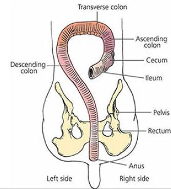
■ FIGURE 12-14 Dorsal view of the dog cecum and colon (large intestine). The dog, a carnivore, has no special arrangement for its ascending colon. The rectum is the pelvic portion of the descending colon that terminates at the anus.
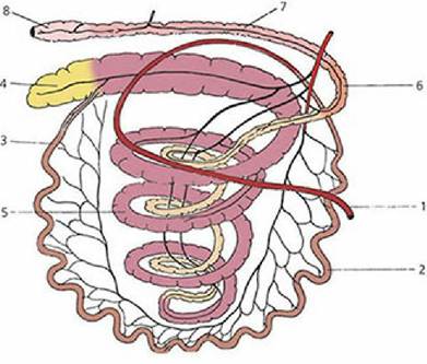
■ FIGURE 12-15 Schematic representation of the intestinal tract of the pig. 1, Rectum; 2, cecum; 3, ileum; 4, ansa spiralis (coiled colon); 5, descending colon; 6, transverse colon; 7, second curve of duodenum; 8, jejunum. (From Engel HH, St Clair LE. Anatomy. In: Leman AD, et al., eds. Diseases of Swine. 6th edn. Ames, IA: Iowa State University Press, 1986.)
In the horse the cecum is a large, comma-shaped structure that extends from the pelvic inlet to the abdominal floor, with its tip just behind the diaphragm (Figure 12-17). It is mainly located on the right side of the horse. The ventral colon continues cranially from the base of the cecum, which is near the pelvic inlet on the right side, to the diaphragm, where it turns caudad and returns to the pelvic inlet. Another turn is made cranially and continues as the dorsal colon, located above the ventral colon. The ventral and dorsal colons can be described as double horseshoes because one seems to be on top of the other. A turn is made at the diaphragm, and the dorsal colon continues for a short distance and joins the transverse colon, which is directed toward the left side of the horse. The descending colon in the horse is called the small colon.
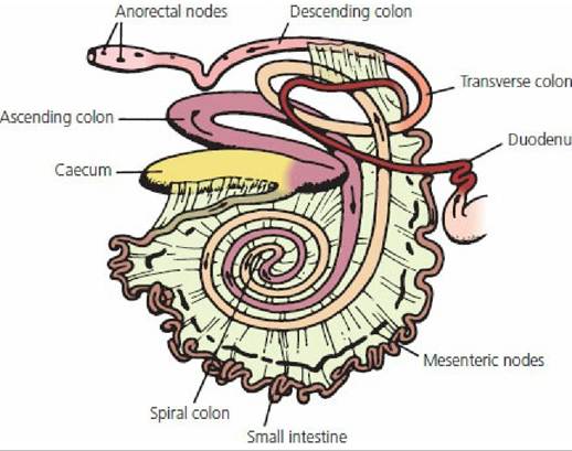
■ FIGURE 12-16 Gastrointestinal tract of the cow showing the colic spiral (ansa spiralis). (From Dyce KM, Wensing CJG. Essentials of Bovine Anatomy. Philadelphia, PA: Lea & Febiger, 1971.)
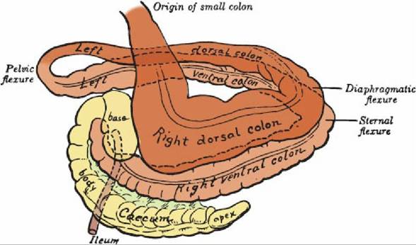
■ FIGURE 12-17 Schematic representation of the cecum and colon of the horse. (From Getty R. Sisson and Grossman’s Anatomy of the Domestic Animals. 5th edn. Philadelphia, PA: WB Saunders, 1975∙)
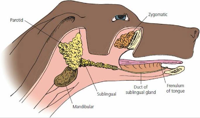
■ FIGURE 12-18 Location of salivary glands in the dog. They are paired glands and only those on the right side are shown. The right mandible has been removed to show the sublingual salivary gland and its duct. The duct empties on a small papilla located near the rostral end of the frenulum (midventral fold of the tongue).
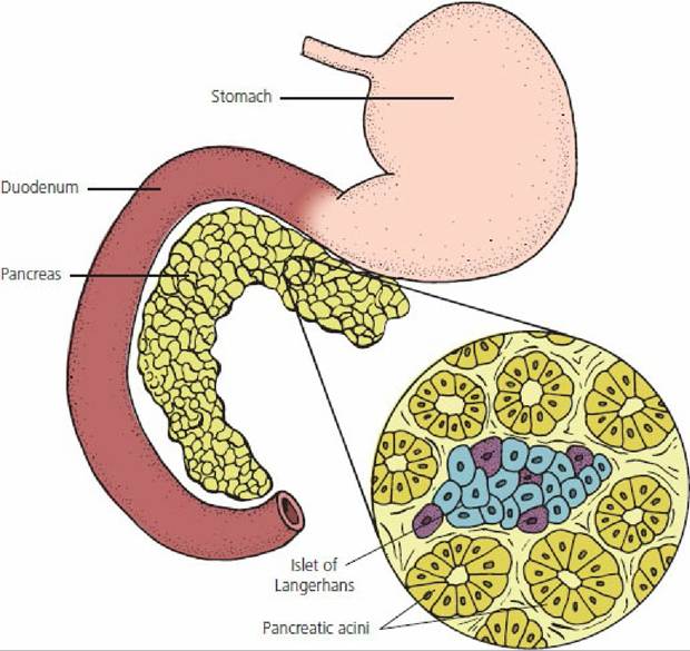
■ FIGURE 12-19 Location of the pancreas and its general appearance. The pancreas is always located near the first part of the duodenum and appears as an elongated gland of loosely connected aggregated nodules. The inset from the pancreas shows an islet of Langerhans (endocrine) situated among a number of pancreatic acini, the exocrine (digestive secretions) portion.
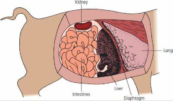
■ FIGURE 12-20 The pig liver and its location relative to other organs. Because of the large amount of interlobular connective tissue, the lobules are mapped out sharply. For this reason, the liver is much less friable (easily broken) than that of other animals.
The cecum and parts of the colon of the pig and horse are sacculated as a result of the presence of longitudinal bands of muscle. The sacculations, called haustra, seem to act as buckets. By accommodating extra volume, they can help to prolong the retention,of contents, thus allowing more time for microbial digestion (see Figures 12-15.and 12-17).
The descending colon terminates at the anus. The part of the descending colon located within the pelvis is known as the rectum. It is relatively dilatable and serves to store feces prior to its expulsion.
The anus is the junction of the terminal part of the digestive tract with the skin. It closes by means of a muscular sphincter composed of smooth and striated muscle fibers.
■