THE SKELETON AND JOINTS
The skeleton is reduced to the bones of the principal digits (III and IV) together with vestiges of those of the flanking ones (II and V) (Figure 30-4). Although the principal metacarpal elements are fused to form a single cannon bone, this divides at its lower end into separate articular trochleae for the two proximal phalanges.
All more distal bones are duplicated. Vestigial structures include the short, rodlike fifth metacarpal bone in articulation with the upper end of the cannon bone (see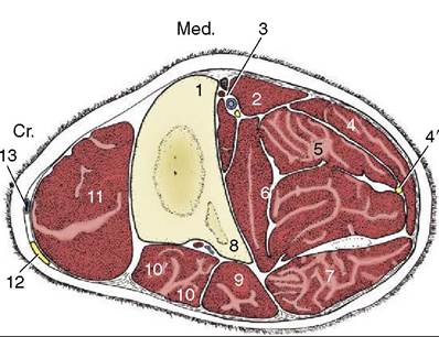
Figure 30-3 Transverse section of the middle of the bovine left forearm. 1, Radius; 2, flexor carpi radialis; 3, median vessels and nerve; 4, flexor carpi ulnaris; 4’, ulnar nerve; 5, superficial digital flexor; 6, deep digital flexor; 7, ulnaris lateralis; 8, ulna; 9, lateral digital extensor; 10, 10', common digital extensor; 11, extensor carpi radialis; 12, superficial branch of radial nerve; 13, cephalic vein.
Figure 2-48) and phalangeal rudiments isolated within the dewclaws.
The cannon bone is compressed from front to back and expanded to the sides at each end. A dorsal axial groove (presenting a vascular foramen at each end) and an incomplete internal septum (visible in radiographs) attest to the composite origin of the bone (Figure 30-9, BZ4). The proximal and middle phalanges are broadly alike, although the former are about twice the length of the latter. All four of these bones present proximopal- mar tubercles, paired on the proximal phalanges and single and abaxial on the middle ones. Each has a distal surface that is grooved sagittally to fit the bifaceted surface of the bone with which it articulates. The distal phalanx is shaped like the hoof in which it is lodged and presents articular, axial, abaxial, and sole surfaces (Figure 30-5).
The extensor process is the highest point, and from it a crest runs to the apex of the bone, dividing the axial and abaxial surfaces. These surfaces are separated caudally by a thick transverse tubercle (Figure 30-5Z4) to which the deep flexor tendon attaches. Apart from the articular surface, the exterior displays numerous vascular foramina most conspicuously on the axial aspect of the extensor process and at the palmar end of the abaxial surface. (The proximal and distal sesamoid bones are described with the joints.)As in the horse, the articulations linking the metacarpal and digital bones are commonly known as the fetlock, pastern, and coffin joints. The fetlock joint, the first duplicated joint of the limb, is slightly overextended when the animal stands at rest (Figure 30-6Z3). Its movements are confined to flexion and extension by reciprocally keeled and grooved articular surfaces and by strong collateral ligaments. The axial (interdigital) collateral ligaments of both joints have a common origin in the intertrochlear notch of the metacarpal bone (see Figure 30-4). The phalangeal articular surfaces are complemented on their palmar aspect by a row of four (proximal) sesamoid bones embedded within a continuous fibrocartilaginous bridge and joined by the interosseous muscle. These sesamoids are additionally secured by collateral and a complex suite of distal sesa- moidean ligaments. The collateral sesamoidean ligaments connect each abaxial sesamoid to the metacarpal bone and proximal phalanx. The ligaments that arise from the distal surfaces pass to the prominent tubercles on the proximopalmar aspect of the related phalanges, crossing in passage to their destinations (cruciate sesa- moidean ligaments); fibers of the axial pair also cross the interdigital space (interdigital phalangosesamoid- ean ligaments; (Figure 30-7Z10). Since the joints enjoy great mobility, the capsules are large; each extends proximally as a dorsal pouch between the metacarpal bone and extensor tendons and as a palmar pouch between the bone and the interosseous muscle (Figure 30-9Z9,9').
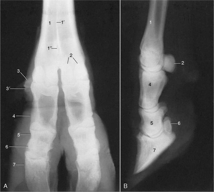
Figure 30-4 Dorsopalmar (A) and Iateromedial (B) radiographs of the bovine foot.
1, Metacarpal bone; Γ, median septum; 1", distal metacarpal canal; 2, proximal sesamoid bones; 3, dewclaw; 3', rudimentary phalanx within dewclaw; 4, proximal phalanx; 5, middle phalanx; 6, navicular bone; 7, distal phalanx.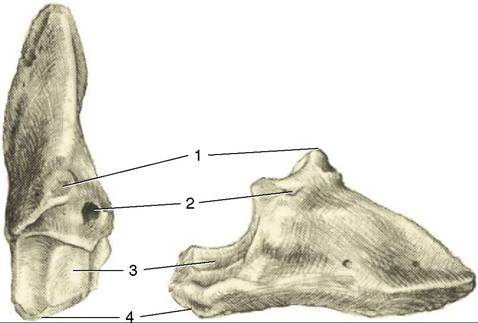
Figure 30-5 The distal phalanx looking distally (left), and axial surface (right). 1, Extensor process; 2, axial foramen for the principal artery to the hoof; 3, articular surface; 4, tubercle on which the deep digital flexor attaches.
Although both may be punctured, the larger palmar pouch is reached more easily; entry is made from the side, about 2 or 3 cm proximal to the joint space. Communication between the paired capsules allows infection or injected material to travel from one joint to the other.
The less mobile pastern joints also allow only flexion and extension. Each joint is supported by one pair of collateral ligaments; the axial one is better developed, presumably to resist the toes spreading apart under the body weight. An additional axial ligament that extends to the distal phalanx is given the same interpretation. The joint obtains further support from a Abrocartilage that extends the palmar border of the articular surface of the middle phalanx and from three palmar ligaments (Figure 30-9, A). The capsules of the two pastern joints are separate. Each forms dorsal and ventral pouches against the proximal phalanx; the dorsal one is said to be accessible to puncture from the side.
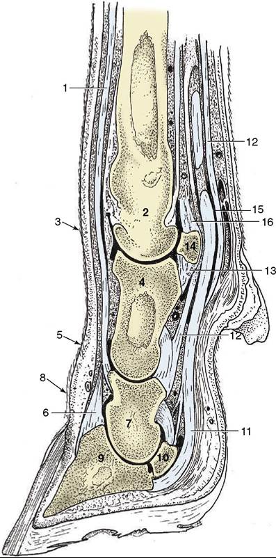
Figure 30-6 Sagittal section of the bovine foot, splitting the lateral digit. 1, Lateral digital extensor; 2, metacarpal bone; 3, fetlock joint; 4, proximal phalanx; 5, pastern joint; 6, common digital extensor; 7, middle phalanx; 8, coffin joint; 9, distal phalanx; 10, navicular bone; 11, deep digital flexor; 12, superficial flexor; 13, distal sesamoidean ligaments; 14, proximal sesamoid bone; 15, digital sheath; 16, interosseous.
The coffin joint resembles the pastern in conformation and in the possession of collateral ligaments. It is entirely within the hoof, and because the small dorsal and ventral pouches barely reach beyond the coronet, puncture is difficult (see Figures 30-6 and 30-9). The distal articular surface is enlarged by the navicular bone located about 2 cm within the hoof (when measured abaxially); its other end is above the axial wall of the hoof, which is lower. The bone is mainly related to the middle phalanx and is held in place by a complex set of distal and collateral ligaments; these pass to the adjacent phalanges and resist overextension. An elastic ligament spanning the axial surface of the joint prompts recollection of the ligament that retracts the claw in cats but appears not to have a comparable function. Interdigital ligaments are also present to prevent splaying of the digits. One connects the axial surfaces of the proximal phalanges (see Figure 30-7), and a second crosses the interdigital space level with the navicular bones, where it is related to the interdigital bridge of skin.