THE SKULL
The salient features of the skull are the large orbits placed between the bulbous cranium and the pyramidal
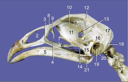
Figure 37-10 Skull of chicken.
1, Premaxilla; 2, nasal aperture; 3, maxilla; 4, jugal arch; 5, mandible; 6, palatine bone; 7, vomer; 8, nasal bone; 9, lacrimal bone; 10, orbit; 11, interorbital septum; 12, frontal bone; 13, optic foramen; 14, pterygoid bone; 15, quadrate bone; 16, temporal bone; 17, parietal bone; 18, occipital bone; 19, tympanic cavity with cochlear and vestibular windows; 20, sphenoid bone; 21, articular bone.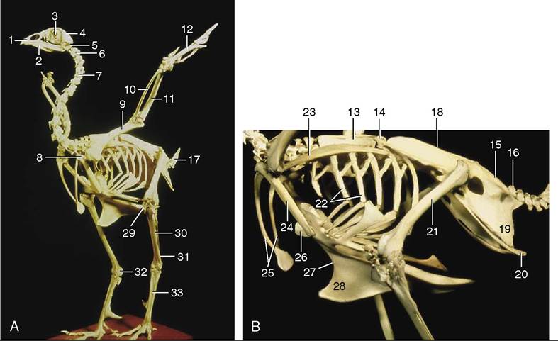
Figure 37-9 Skeleton of a chicken. 1, Facial part of skull; 2, mandible (bones of the hyobranchial apparatus are shown protruding below the mandible); 3, orbit and sclerotic ring of eyeball; 4, cranium; 5, atlas; 6, axis; 7, cervical vertebrae; 8, shoulder joint; 9, humerus; 10, radius; 11, ulna; 12, hand (manus); 13, notarium; 14, free thoracic vertebra; 15, synsacrum; 16, caudal vertebrae; 17, pygostyle; 18, ilium; 19, ischium; 20, pubis; 21, femur; 22, ribs; 23, scapula; 24, coracoid bone; 25, fused clavicles; 26, manubrium sterni; 27, sternum; 28, keel; 29, patella; 30, fibula; 31, tibiotarsus; 32, sesamoid bone (ossified tibial cartilage) in hock joint; 33, tarsometatarsus.
face (Figure 37-10). The mandible is flat and adds only marginally to the height of the head. The enormous eyes have displaced the bones found between the orbits in most mammalian skulls and have reduced others to a thin median plate (interorbital septum; Figure 37-10/11). Several cranial bones consist of two plates separated by spongy bone; they are thus thicker than would be supposed and give the impression that the cranial cavity is greater than it is.
The occipital bone encloses the foramen magnum. A single occipital condyle immediately ventral to this articulates with the atlas, forming a joint that enables birds to rotate the head on the vertebral column to a much greater extent than is allowed to mammals. The semispherical depression in the lower part of the lateral cranial wall is the tympanic cavity (Figure 37-10/19). Its rim bounds the external acoustic meatus, which is closed by the tympanic membrane in life. Cochlear and vestibular windows in the depth of the depression lead into the inner ear.The facial part of the skull is formed principally by the nasal and premaxillary bones that surround the large nasal aperture (Figure 37-10/2). The nasal bone is dorsal, and in many birds, for example, in the psittacine species, it makes a flexible cartilaginous connection with the frontal bone, which permits the upper jaw to be raised as the mandible is depressed. The maxilla below the nasal aperture is small and is connected to the mandibular joint by the long and thin jugal arch (Figure 37-10/4), the homologue of the mammalian zygomatic arch. The palatine bones (Figure 37-10/d) are caudally directed rods connecting the premaxillae with the pterygoid bones ventral to the orbits. Thus the osseous partition between the nasal and oral cavities exists only rostrally, where it is formed by the palatine processes of the premaxillae.
The mandible (Figure 37-10/5) consists of two thin bones fused rostrally where they are covered by the lower beak. Caudally, the mandible is connected to the skull between the orbit and the external acoustic meatus by the articular and quadrate bones (Figure 37-10/15,21), which are elements that correspond to mammalian middle ear ossicles, the malleus and incus. The quadrate bone is connected to the jugal arch, and, by interposition of the pterygoid, to the rodlike palatine bone. In birds with a craniofacial hinge, depression of the lower jaw rotates the quadrate bone, which pushes the jugal arch and palatine bone rostrally, thus elevating the upper jaw (craniokinesis). In budgerigar and parrots this elastic hinge is replaced by an articular craniofacial joint, which allows even more flexibility of movement.