THE SPLEEN
The spleen is contained within the left cranial part of the abdomen where it is joined to the greater curvature of the stomach by inclusion within the greater omentum. This helps fix its position, which cannot be defined with great precision as it is dependent on the degree of filling of the stomach and on its own blood content.
The basic form is very dissimilar in the various domestic species, being dumbbell-shaped in the dog and cat, straplike in the pig, a broader oblong shape in cattle, and falciform in the horse (Figure 7-61). Its capsule extends trabeculae into the interior. In some species (carnivores) the capsule and trabeculae are very muscular, in others (ruminants) much less so; these differences determine the extent of the physiological variation in size that may occur. When relaxed, the spleen of the dog and cat increases severalfold from its contracted state; it is therefore particularly effective as a reservoir from which the cell content of the circulation may be recruited in times of stress.The soft tissue contained within the supporting framework is divided between red and white pulp; the former consists of spaces in series with the blood vessels and is occupied by a concentration of the cellular elements of the blood. The white pulp, which is divided into foci that are usually just visible to the naked eye, is formed of lymph nodules within a supporting reticuloendothelial framework. This tissue has the usual lymphogenic and phagocytic properties.
The functions of the spleen are blood storage, the removal of particulate matter from the circulation, the destruction of worn-out erythrocytes, and the produc-
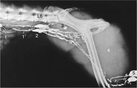
Figure 7-60 Lymphangiogram of the canine lumbar area, pelvis, and thigh. 1, Lumbar aortic lymph node; 1', lumbar trunks; 2, medial iliac nodes; 3, hypogastric node; 4, thigh muscles; 5, popliteal nodes; L6, sixth lumbar vertebra.
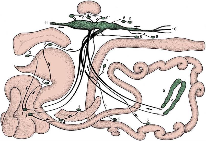
Figure 7-59 Lymph drainage from the organs in the canine abdominal and pelvic cavities (schematized).
1, 1', Right and left hepatic nodes; 2, gastric node; 3, splenic nodes; 4, pancreaticoduodenal nodes; 5, jejunal nodes; 6, right colic node; 7, middle colic node; 8, caudal mesenteric nodes; 9, lumbar aortic nodes; 9', renal nodes; 10, efferents from the iliosacral region; 11, continuation of cisterna chyli as thoracic duct.tion of lymphocytes. The first role is familiar to all who have experienced a “stitch,” the pain that sometimes accompanies physical stress and is associated with contraction of the splenic capsule.
The spleen is supplied by the splenic artery, a branch of the celiac artery that is generously sized in relation to the organ (see Figure 3-39/4). The venous drainage through the splenic vein leads to the portal vein (see Figure 3-50/1,2). Important specific features in the arrangement of these vessels exist. The artery and vein may pass undivided through a confined hilus (ruminants; Figure 7-61, B); run the length of the organ, detaching branches at intervals (horse, pig; Figure 7-61, A); or divide as they approach the spleen into branches that vascularize splenic compartments that are normally independent, although they do communicate (dog, cat; Figure 7-61, C). The lymph vessels found in the capsule and trabeculae do not extend into the pulp. The sympathetic and parasympathetic nerves approach with the artery.
The spleen develops from a mesodermal condensation within the dorsal mesogastrium (which becomes the greater omentum) (see Figure 3-65/6). The part of the sheet intervening between the stomach and the spleen may be specifically distinguished as the gastrosplenic ligament.