THE STIFLE, LEG, AND HOCK
The stifle joint resembles that of the horse in possessing three patellar ligaments and an asymmetrical trochlea (Figure 31-4, B). The patella, patellar ligaments, and tibial tuberosity can be palpated on the cranial surface; two palpable “dimples” at the proximal end of the tuberosity separate and conveniently identify the three ligaments.
The prominent femoral epicondyle, collateral ligament (and its attachment to the rudimentary fibula; Figure 31-4, A/9), and, more cranially, the common origin of the long digital extensor and peroneus tertius (Figure 31-4/5) are palpable on the lateral aspect. As in the horse, the intermediate patellar ligament, the patella, a medial fibrocartilage, and the medial patellar ligament combine to form a loop that passes over the expanded proximal end of the medial ridge (Figure 31-4, B/11) of the femoral trochlea. Although relatively little muscular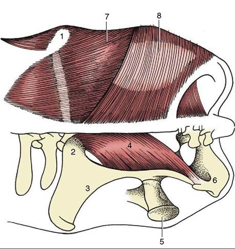
Figure 31-1 Dorsal view of the bovine croup; the muscles on the left side have been removed. 1, Coxal tuber; 2, sacral tuber; 3, ilium; 4, sacrosciatic ligament; 5, greater trochanter of femur; 6, ischial tuber; 7, gluteus medius; 8, biceps.
effort keeps the loop in place (which prevents flexion of the stifle), the mechanism is by no means as efficient as that of the horse, in which the stifle can be fully locked. Lateral and medial luxations of the patella are occasionally reported. Dorsal dislocation, better described as fixation, is more common; indeed it is relatively prevalent among working bullocks of the Indian subcontinent. The condition is usually intermittent and, if not relieved spontaneously, may be treated by section of the medial patellar ligament.
The femoropatellar and medial femorotibial joint cavities always communicate, but the lateral femoro- tibial joint does not communicate with either of the other two.
Two puncture sites are therefore in use. One, between the medial and intermediate patellar ligaments a short distance proximal to the tibia, gives access to the femoropatellar space; the other, in the extensor groove of the tibia, cranial to the common tendon of the long digital extensor and peroneus tertius, provides access to the lateral femorotibial compartment.The tibia is the only weight-bearing bone of the leg (crus). Its medial surface, including the prominent medial malleolus, is subcutaneous; the remaining surfaces are covered by muscle (see Figure 31-6). The distal articular surface (cochlea) presents two sagittal grooves separated by a ridge; each groove is bounded externally by the corresponding malleolus. The fibula is much reduced. A proximal rudiment, generally drawn into a distal point, is fused with the lateral condyle of the tibia
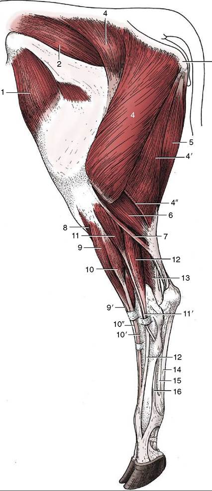
Figure 31-2 Muscles of the bovine left hindlimb; lateral view. 1, Tensor fasciae latae; 2, gluteus medius; 3, ischial tuber; 4, 4', 4'', biceps, transected at 4''; 5, semitendinosus; 6, lateral head of gastrocnemius; 7, rudimentary soleus; 8, tibialis cranialis; 9, 9', peroneus tertius; 10, 10', 10", long digital extensor; 11, 11', peroneus longus; 12, lateral digital extensor; 13, lateral digital flexor; 14, tendon of superficial digital flexor; 15, combined tendon of deep digital flexors; 16, interosseous.
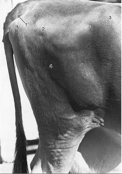
Figure 31-3 Right bovine thigh. 1, Ischial tuber; Γ, sacro- tuberous part of sacrosciatic ligament; 2, greater trochanter of femur; 3, coxal tuber; 4, biceps; 5, lateral vastus; 6, patella; 7, flank fold.
and receives the lateral collateral ligament of the stifle. The distal rudiment is a separate (and palpable) quadrilateral bone (lateral malleolus; Figure 31-5/2) that articulates securely with the tibia by means of an interlocking spike and groove.
It also takes part in the formation of the hock joint.The tarsal skeleton is formed by the following elements: calcaneus and talus in the proximal row; fused central and fourth bones in the intermediate row; and fused second and third bones and a small independent first bone in the distal row (see Figure 2-60). In marked contrast to that of the horse, the talus carries a trochlea at each end (as in artiodactyls generally; Figure 31- 5/4',4"). The proximal trochlea articulates with the tibial cochlea and malleolar bone, forming the tarsocrural joint; the distal trochlea articulates with the calcaneus behind and the fused central and fourth tarsal bones distally, forming the proximal intertarsal joint. Both joints allow flexion and extension, the principal movements at the hock; the proximal joint has the greater excursions. The calcaneus, more slender than the equine bone, has an additional articulation with the lateral malleolus. The tuber calcanei (point of hock) is slightly expanded. The combined central and fourth tarsals (Figure 31-5/5) span the breadth of the hock. The part provided by the fourth tarsal extends into the distal row and articulates with the metatarsal bone. It is related to the fused second and third bones on its medial side. The small first tarsal lies on the plantar aspect of the joint. The surfaces of the distal elements that concur in the formation of the distal intertarsal and tarsometatarsal joints are relatively flat and permit minimal movement. A small discoid sesamoid bone on the plantar surface of the metatarsal bone is embedded in the proximal part of the interosseous (Figure 31-5/7).
Few of the many ligaments are individually important. The joint is supported on each side by collateral ligaments whose long components may be palpated in their full extents from the respective malleolus to the metatarsus. The long plantar ligament (palpable on the plantaromedial aspect) follows the plantar border of the calcaneus and extends beyond this to the metatarsus; it unites the bones on the plantar aspect that would otherwise be pulled apart by the powerful muscles attaching on the point of the hock.
The tarsocrural and proximal intertarsal articulations share a common and relatively capacious cavity. When enlarged, the capsule pouches noticeably on the dorsomedial aspect of the hock, medial to the tibialis cranialis tendon and directly distal to the medial malleolus. It can be punctured more safely than in the horse because the pouch is not overlain by a vein. The other joints are rarely of clinical concern.
The conformation of the hindlimb, particularly the hock, is important in the selection of animals for breeding. The points of the hock should be vertically below the ischial tubers in both lateral and caudal views. If they are too close the animal is said to be “cow-hocked,” and its feet assume a wide stance. An adaptation to an overlarge udder is one cause of an exaggerated approximation of the points of the hocks. (The opposite bowlegged conformation brings the feet close together.) The normal angle of the hock joint (viewed from the side) is about 140°, which gives the metatarsus a slightly forward inclination. When the angle is noticeably smaller, the hock sinks and the animal is said to be “sickle-hocked”; when it exceeds the normal, the animal is said to be “straight-hocked,” a defect that may lead to “weak pasterns” because of the reduced angle at the fetlock joint. Abnormal postures of the hock cause faulty footing and risk damage to the tendons and synovial structures of the digits.
The muscles of the leg are divided into the usual craniolateral and caudal groups. Among the former, the tibialis cranialis and peroneus tertius broadly resemble those of the horse (Figure 31-2/8,9); the peroneus tertius, though largely tendinous, is yet significantly fleshier than its equine equivalent. The long digital extensor resembles the common extensor of the fore-
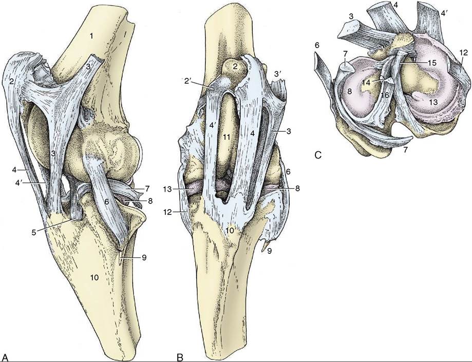
Figure 31-4 The left bovine stifle joint. A, Lateral view. B, Cranial view. C, The menisci and ligaments attaching on the proximal end of the left tibia.
1, Femur; 2, patella; 2', Abrocartilage of patella; 3, lateral patellar ligament; 3', attachment of biceps; 4, intermediate patellar ligament; 4', medial patellar ligament; 5, combined tendon of long digital extensor and peroneus tertius; 6, lateral collateral ligament; 7, tendon of popliteus; 8, lateral meniscus; 9, fibula; 10, tibia; 10', tibial tuberosity; 11, medial ridge of femoral trochlea; 12, medial collateral ligament; 13, medial meniscus; 14, cranial cruciate ligament; 15, caudal cruciate ligament; 16, meniscofemoral ligament.limb in possessing two bellies: one supplies the tendon proper to the medial digit, while the tendon of a second, smaller one splits to reach both digits. There is also a lateral extensor (Figure 31-2/72), proper to the lateral digit. All extensor tendons are of necessity held in place by (two) stout, palpable retention bands where they descend over the flexor surface of the hock; equally necessarily, synovial sheaths protect them here. The proximal retinaculum is easily palpated even in heavy, thick-skinned cows. The group is completed by a peroneus longus muscle (Figure 31-2/77) that arises near the lateral collateral ligament of the stifle and descends on the lateral side of the leg. It then crosses over the tendon of the lateral digital extensor to wind around to the plantar aspect of the hock where it inserts. Some inward rotation of the foot is produced by its contraction.
The gastrocnemius (Figure 31-2/b) arises by twin heads from the caudal surface of the femur and forms a muscular swelling at the upper end of the leg before narrowing abruptly to the strong tendon that inserts on the point of the hock.
The superficial digital flexor, though more muscular than that of the horse, is very tendinous and relatively inextensible (Figure 31-6/74). It arises between the heads of the gastrocnemius, winds around the medial surface of that muscle’s tendon, and spreads to cap the point of the hock. The edges of the cap attach here, but the bulk of the tendon continues down the plantar surface into the foot.
The crural segment, acting in concert with the peroneus tertius, links the movements of the stifle and hock joints. (This needs to be kept in mind when attempting to correct the relatively common breech position of a fetus that presents the tail and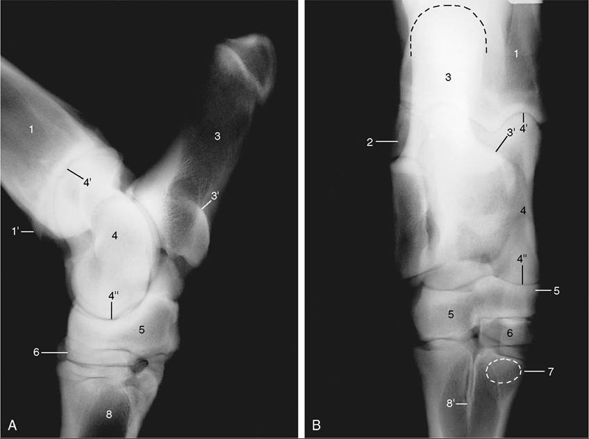
Figure 31-5 Lateral (A) and dorsoplantar (B) radiographs of the bovine hock. 1, Tibia; 1', medial malleolus; 2, lateral malleolus (distal end of fibula); 3, calcaneus; 3', sustentaculum tali; 4, talus; 4', 4", proximal and distal trochlea of talus; 5, fused central and fourth tarsal bones; 6, fused second and third tarsal bones, in B superimposed on small first tarsal bone (not labeled); 7, position of sesamoid bone in interosseous; 8, metatarsal bone; 8', median septum.
flexed hocks.) An extensive subtendinous (calcanean) bursa protects the tendon both where it wraps around the gastrocnemius and again over the point of the hock. Occasionally a subcutaneous bursa (hygroma) develops over the tendon here.
The gastrocnemius and superficial flexor are in a continuous (reflex) state of contraction in calves with “spastic paresis.” In these animals the hock and stifle are maximally extended, and the affected limb is used stiffly with only the toes of the hoofs touching the ground (Figure 31-7). Section of the tendons (or of the [tibial] nerve branches to the gastrocnemius) gives relief. Although there is no proof of inheritance, it is generally agreed that it is unwise to breed from affected animals even after surgical “cure.”
The deep digital flexor (Figure 31-6/9) has three heads. Two come together in the leg to form a thick tendon that passes over the plantar surface of the hock medial to the calcaneus and is protected by the tarsal synovial sheath. The tendon is bound down by the flexor retinaculum and other deep fasciae so that, when distended, the sheath bulges only at its ends, proximal
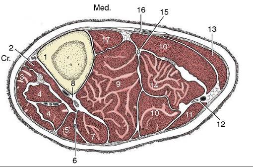
Figure 31-6 Transverse section of the left bovine leg. 1, Tibia; 2, tibialis cranialis; 3, peroneus tertius; 4, long digital extensor; 5, peroneus longus; 6, peroneal nerve; 7, lateral digital extensor; 8, cranial tibial vessels; 9, deep digital flexors; 10,10', lateral and medial heads of gastrocnemius; 11, biceps;
12, caudal cutaneous sural nerve and lateral saphenous vein;
13, semitendinosus; 14, superficial digital flexor; 15, tibial nerve; 16, saphenous vessels and nerve; 17, popliteus.
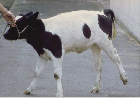
Figure 31-7 Calf with spastic paresis.
and distal to the joint. The thin tendon of the third head tunnels through the dense medial tarsal fascia, within its own synovial investment, to join the major tendon in the metatarsus. The popliteus has no special features.
Most locomotor and cutaneous structures of the hindfoot are very similar to their forelimb counterparts and need not be described. However, the metatarsal bone is noticeably longer than the metacarpal and is quadrilateral in transverse section, which gives the hind cannon a deeper appearance in lateral view (see Figure 31-14). The higher incidence of disease in the digits of the hindlimb, especially the lateral one, has not been fully explained.