» The Stomach
The most remarkable feature of the stomach is its small size (5- to 15-L capacity) in relation to the animal and to the volume of fodder consumed. It is relatively larger in the unweaned foal.
The equine stomach lies mainly within the left half of the abdomen (Fig. 21.10/2). Like other simple stomachs, it consists of two limbs that meet at a ventral angle. The left limb comprises the fundus (unusually large and often termed the saccus cecus [blind sac] in this species) and the body; the right limb or pyloric part is much narrower and extends across the midline to join the duodenum (Fig. 21.11A). It mostly lies within the rib cage and is inaccessible through the flank or the rectum even when grossly distended. Gross overdistention may be revealed by a raising of the overlying ribs on the left side, which destroys the normal symmetry of the trunk.
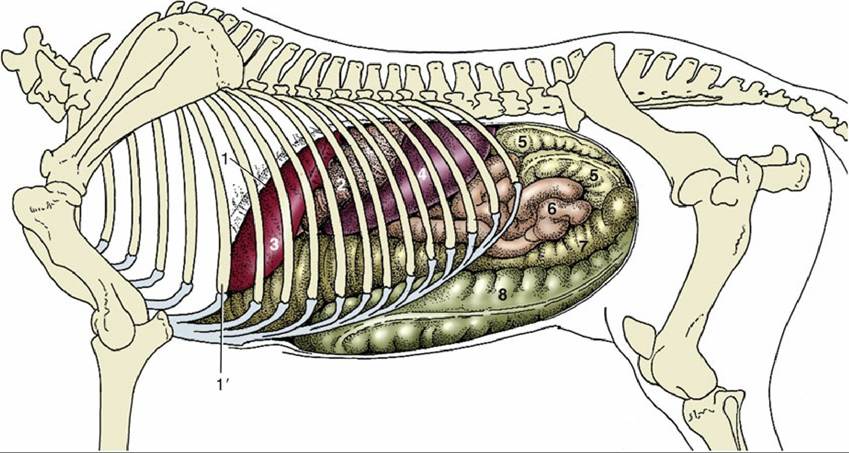
FIG. 21.6 Visceral projections on the left abdominal wall (including the diaphragm). 1, Cut edge of diaphragm; 1', rib 6; 2, stomach; 3, liver; 4, spleen; 5, descending colon (banded); 6, jejunum (smooth); 7, left dorsal colon; 8, left ventral colon.
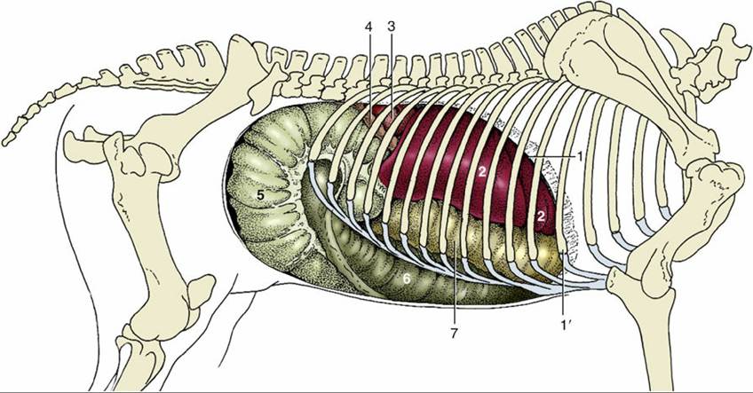
FIG. 21.7 Visceral projections on the right abdominal wall (including the diaphragm). 1, Cut edge of diaphragm; 1', rib 6; 2, liver; 3, right kidney; 4, descending duodenum; 5, body of cecum; 6, right ventral colon; 7, right dorsal colon.
Stomach Surface Projections: When moderately distended, the fundus extends under the upper part of the 15th rib (or thereabouts), and the lowest part of the body reaches the ventral parts of the 9th and 10th ribs. The cardia provides a relatively fixed point, opposite the upper part of the 11th rib, and enlargement after feeding is therefore mainly downward and forward (Fig.
21.6/2).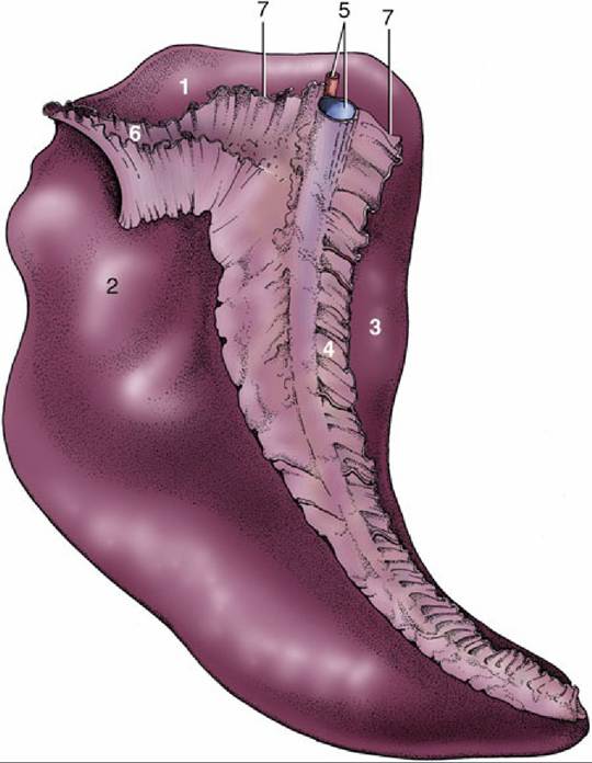
FIG. 21.8 Visceral surface of the spleen. 1, Renal surface; 2, intestinal surface; 3, gastric surface; 4, greater omentum (gastrosplenic ligament); 5, splenic artery and vein; 6, renosplenic ligament; 7, phrenicosplenic ligament.
The cranial surface is directed against the diaphragm above and against the left lobe of the liver more ventrally. The caudal surface faces in the opposite direction and makes contact with various viscera, including coils of small intestine and descending colon dorsally and the dorsal diaphragmatic flexure of the ascending colon ventrally. The left part of the greater curvature is followed by the hilus and adjoining gastric surface of the spleen (see Fig. 21.9).
A stepped edge (margo plicatus; Fig. 21.9/2") divides the interior between a large nonglandular region, occupying the fundus and part of the body, and a glandular region. The nonglandular part resembles the mucosa of the esophagus and is dirty white and harsh to the touch (see Fig. 21.11). The softer glandular region consists of cardiac, proper gastric, and pyloric glandular zones. Although the borders between these zones are ill defined, the zone occupied by the proper gastric glands is somewhat darker and redder than the yellowish cardiac and pyloric zones in the fresh specimen. Both the cardiac and pyloric regions are incidentally parasitized by botfly (Gasterophilus) larvae, which may leave the mucosa densely pocked by small focal ulcerations. These, when semihealed, can be misinterpreted as normal features (Fig. 21.11B).
The cardiac sphincter is exceptionally well developed, and this, coupled with the oblique entrance of the esophagus, is held responsible for the horse's reputed inability to eructate or vomit. However, eructation and vomiting, though rare, is possible. The canal or distal portion of the pyloric part is more muscular than the remainder of the organ and is bounded by proximal and distal thickenings that converge at the lesser curvature. Even when the second of these, the pyloric sphincter, is fully relaxed, the actual exit is remarkably narrow (Fig. 21.9/5).
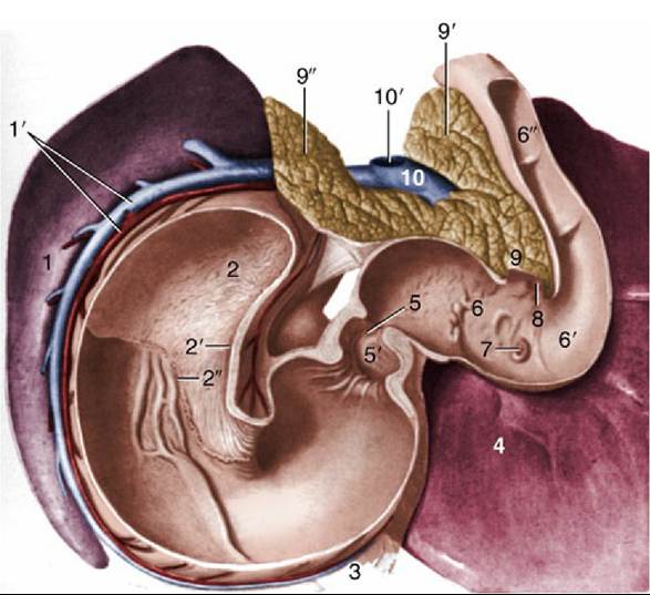
FIG. 21.9 Topography of spleen, stomach, pancreas, and liver, Caudoventral view. 1, Intestinal surface of spleen; 1', splenic artery and vein; 2, fundus (blind sac) of stomach; 2', cardia; 2", margo plicatus; 3, greater omentum; 4, liver; 5, pyloric orifice; 5', pyloric antrum; 6, S-shaped cranial part of duodenum; 6', cranial flexure of duodenum; 6", descending duodenum; 7, major duodenal papilla; 8, minor duodenal papilla; 9, body of pancreas; 9' and 9", left and right lobes of pancreas, respectively; 10, portal vein; 10', stump of cranial mesenteric vein.