The Stomach
Species variation in the gastrointestinal tract is most marked where the stomach is concerned. The stomach of fish- and flesh-eating species (raptors, hawks, ospreys, vultures, and owls) is primarily a storage organ appropriate for the chemical digestion of a soft diet.
In contrast, the stomach of birds with a herbivorous diet is adapted to the mechanical reduction of tougher material through powerful muscular development. Domestic poultry (chicken, geese, and others that are similar) possess stomachs of the second category and exhibit only minor interspecific variation.The stomach of these birds is divided by a constriction (isthmus) into a predominantly glandular proventriculus and a predominantly muscular ventriculus (gizzard) placed one behind the other close to the median plane. The proventriculus is ventrally in contact with the left lobe of the liver. The larger, more caudal gizzard has more extensive contact with the sternum and the lower part of the left lateral abdominal wall. It is exposed when the sternum and abdominal muscles are removed during necropsy (Fig. 37.17).
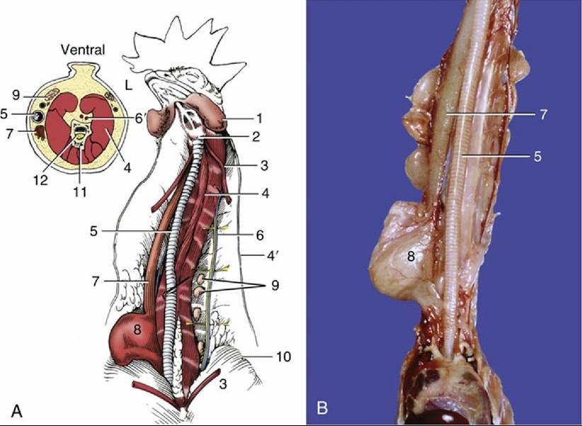
FIG. 37.15 Ventral view of the dissected neck. (A) Schematic; the inset shows a transverse section through the middle of the neck. (B) Detail of neck with crop. L, Left side of the transverse sect; 1, wattle; 2, larynx; 3, sternothyroideus, cut; 4, cervical muscles; 4', cervical nerve; 5, trachea; 6, jugular vein and vagus nerve; 6', internal carotid arteries; 7, esophagus; 8, crop; 9, thymus; Left side of the transverse section 10, pectoralis; 11, vertebra; 12, spinal cord.
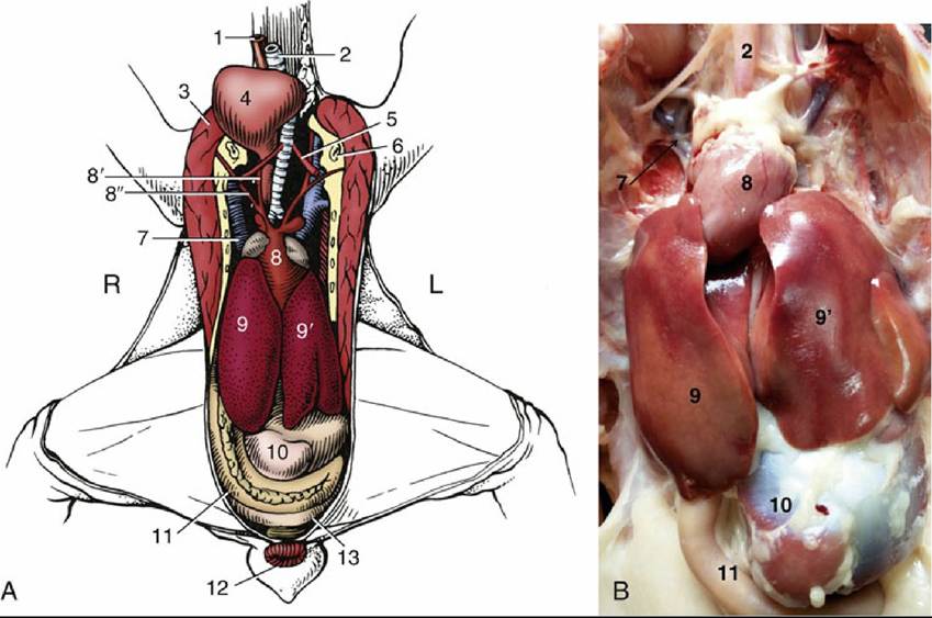
FIG. 37.16 Ventral view of the viscera. (A) Schematic. (B) Viscera after removal of ventral body wall, ventral view.
L, Left; R, right; 1, esophagus; 2, trachea; 3, pectoralis, cut; 4, crop; 5, Sternotrachealis; 6, coracoid bone, cut; 7, right cranial vena cava; 8, heart; 8', common carotid artery; 8", subclavian artery; 9 and 9', right and left lobes of liver, respectively; 10, gizzard (its caudal blind sac); 11, duodenal loop, enclosing pancreas; 12, vent; 13, one of the ceca.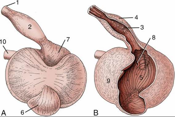
FIG. 37.17 Stomach, ventral surface (A) and opened ventrally (B). 1, Esophagus; 2, proventriculus; 3, papillae; 4, deep proventricular glands, visible on cut surface; 5, lumen of gizzard; 6, caudal blind sac; 7, cranial blind sac; 8, pyloric orifice; 9, cranioventral muscle mass; 10, duodenum.
The proventriculus is spindle shaped and about 4 cm long. Its whitish mucosa, lined with a mucussecreting, columnar epithelium, is clearly demarcated from the more reddish lining of the esophagus (Figs. 37.18 and 37.19). It presents numerous macroscopic elevations (papillae) through which pass the collecting ducts from a thick bed of glands, very visible on the cut surface of the wall. The papillae are so prominent that they may be mistaken for parasitic lesions. There are two kinds of epithelial cells in the glands: oxynticopeptic cells that produce both hydrochloric acid and pepsinogen and cells that produce mucus.
The isthmus is the transition from the glandular stomach to the muscular gizzard. It has no glands in its thinner, less rigid wall. In many parrots the koilin layer from the gizzard extends some way into it.
The ventriculus or gizzard is lens shaped in herbivores, poultry, and waterfowl and is positioned with its convex surfaces facing more or less to the right and left (Fig. 37.17/5). Its interior is elongated, enlarged by cranial and caudal blind sacs, of which the former connects with the proventriculus. The duodenum arises on the right surface, adjacent to the cranial blind sac.
The bulk of the organ consists of two thick masses of muscle that insert on glistening tendinous centers, one on each surface. Thinner muscles cover the blind sacs. The mucous membrane is thin but very tough. It has a cuboidal epithelium and largely consists of tubular glands. Catalyzed by the low pH resulting from the hydrochloric acid from the proventriculus, the secretion of the glands forms a hard cuticle of koilin (a carbohydrate-protein complex). The koilin, a rough plicated layer, is replenished from the glands below as it is worn on the surface. It obtains a yellow-green color from the bile refluxed from the duodenum. In herbivorous and omnivorous birds, powerful contractions of the gizzard crush the food, assisted by ingested grit, which must be provided in the diet (Fig. 37.19). Being radiodense, the grit identifies the gizzard in radiographs. The gizzard is the site of protein digestion.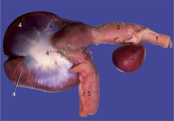
FIG. 37.18 Stomach of chicken. 1, Esophagus; 2, spleen; 3, proventriculus; 4, gizzard with aponeurosis (4'); 5, duodenum.
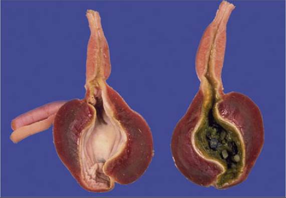
FIG. 37.19 Opened stomach. Note grit inside gizzard (right).
In granivores, psittacine species, and songbirds the gizzard is less muscular because these birds dehusk and crumble their seeds before swallowing. These birds do not always require grit.
Muscular activity moves food back and forth between the proventriculus and gizzard during digestion; the location of the pylorus then enables some of the food that does not require grinding to escape into the duodenum, bypassing the gizzard.