The Structure of Cardiac and Smooth Muscle Differs from That of Skeletal Muscle
As with skeletal muscle, cardiac muscle is striated and contains sarcoplasmic reticulum and myofibrils; the fundamental contractile component is formed by actin and myosin subunits (see Figure 19-1).
Cardiac muscle also contains transverse tubules, but cardiac muscle differs from skeletal muscle in some important ways. The long skeletal muscle fibers are electrically isolated from each other, whereas the shorter cardiac muscle cells are electrically coupled to each other through end-to-end intercalated disks that contain gap junctions. Because gap junctions provide continuity between the cytoplasm of adjacent cells, action potentials can spread from one cardiac muscle cell to another, across these intercalated disks, without the need for chemical neurotransmission to each cell. The cardiac muscle cells can also possess branchlike extensions that form similar connections with some of their parallel neighbors. In fact, as explained in Chapter 19, action potentials arise spontaneously in specialized cardiac muscle cells and then spread throughout a large population of cardiac muscle cells as if they were a functional syncytium. This can result in coordinated contraction of a large region of the heart muscle. The frequency of such action potentials and the force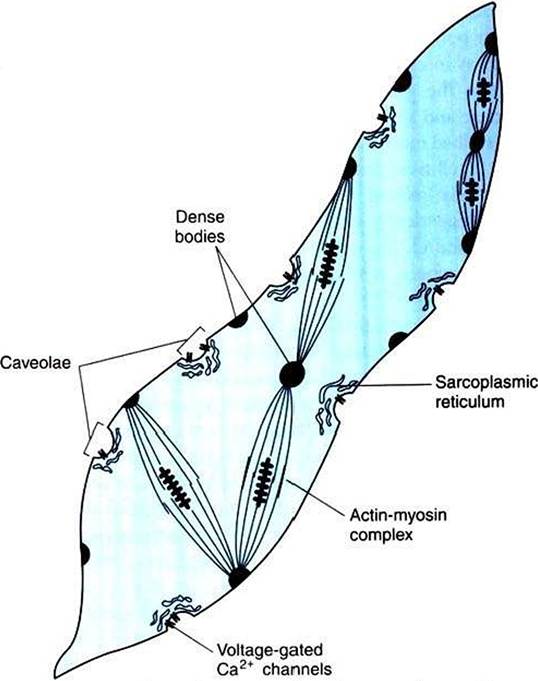
FIGURE 6-7 General organization of a smooth muscle cell.T tubules are absent, and the sarcoplasmic reticulum is poorly Jeveloped-Transmembrane diffusion of extracellular Ca2*, through voltage-gated Ca2* channels in caveolae, plays an important role in initiating contraction. Actin and myosin are present, with actin anchored to dense bodies. Activating the actin-myosin complex can change the cell's shape. (Modified from Guyton AC, Hall JE: Textbook of medical physiology, ed 11, Philadelphia, 2006, Saunders.)
of the resulting contraction are influenced by the autonomic nervous system, but such innervation is not necessary for action potential genesis.
Smooth muscle cells, as with cardiac myocytes, are smaller and shorter than skeletal muscle cells. They do not contain T tubules, and their sarcoplasmic reticulum is poorly developed (Figure 6-7). These cells rely primarily on the transmembrane diffusion of Cinflux is achieved, on membrane depolarization, through activation of voltage-gated Ca2 ‘ channels located in shallow depressions of the membrane (caveolae) (see Figure 6-7). Calcium channel blockers interfere with this process and can relax smooth muscle in arterial walls, which dilates arteries and lowers blood pressure. Contraction is terminated in many smooth muscle cells primarily by Ca2* transport back into the extracellular space, which is a fairly slow process.
The mechanism by which Ca2' induces actin-myosin cross-bridge cycling in smooth muscle differs from that in skeletal and cardiac muscle (see Figure I-17). In skeletal and cardiac muscle, cross-bridge cycling relies primarily on the Ca2 -induced removal of the tropomyosin block of the actin- binding site. In smooth muscle, cycling relies on a Ca2'- induced increase in the AFPase activity of the myosin head, another slow process.