The Structure of the Heart
The thick middle layer of the wall (myocardium) is composed of cardiac muscle, which is a variety of striated muscle peculiar to this organ. It is covered externally by the visceral pericardium (epicardium) and internally by the endocardium, a thin smooth-surfaced layer continuous with the lining of the blood vessels.
The atrial and ventricular parts of the muscle are separated by a fibrous skeleton that is formed
mainly by the conjunction of the rings that encircle the four heart orifices. The skeleton contains islands of fibrocartilage in which nodules of bone (ossa cordis) may develop (Fig. 7.12/5). Although these bones appear precociously in the hearts of cattle, they are not confined to this species, as is sometimes suggested. Near the entrance of the coronary sinus, the fibrous skeleton allows passage to the atrioventricular bundle, which conducts the impulse to contract and constitutes the only direct connection between the atrial and ventricular muscles. Delicate extensions of the fibrous tissue also provide the cores of the cusps of the various valves.
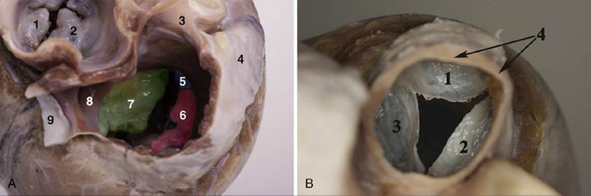
FIG. 7.14 (A) Atrioventricular openings of the dog (dorsal view): 1, Parietal cusp of the left
atrioventricular valve; 2, septal cusp of the left atrioventricular valve; 3, cranial vena cava; 4, right auricle; 5, angular cusp of the right atrioventricular valve; 6, parietal cusp of the right atrioventricular valve; 7, septal cusp of the right atrioventricular valve; 8, opening of the coronary sinus; 9, caudal vena cava. (B) Semilunar valves of the pulmonary trunk: 1, Intermediate semilunar valve; 2, right semilunar valve; 3, left
semilunar valve; 4, pulmonary trunk.
The atrial muscle is thin—indeed, the auricular wall may be translucent between the pectinate ridges. It is arranged in superficial and deep bundles; some of the former are common to both atria, but the remainder, and all of the deep bundles, are confined to one.
It has been postulated that the fascicles that surround the various systemic and pulmonary venous inlets act as throttles to oppose reflux of blood into the veins during atrial systole.The much thicker ventricular muscle is also arranged in superficial and deep bundles. Some superficial bundles coil around both chambers, utilizing the septum to complete a figure-of-eight course. Others, like the deeper bundles, encircle only the one chamber. The arrangement of the muscles and their contraction mechanisms are actually very complicated.
The inherent rhythm of the heart is controlled by a pacemaker, a small, richly innervated sinoatrial node of modified cardiac fibers (nodal myofibers) (Fig. 7.16A). This node, which is not apparent to the naked eye, lies below the epicardium of the right atrial wall ventral to the cranial caval opening (see Fig. 7.3/11). With each heart cycle a wave of excitation arises in the sinoatrial node and spreads throughout the atrial muscle to reach the atrioventricular node (Figs. 7.15 and 7.16B and C). In ungulates, specialized conductive tissue is present subendocardially in the atrium, mainly on the pectinate muscle. From the atrioventricular node an excitatory stimulus passes rapidly throughout the whole ventricular myocardium via the atrioventricular bundle, which is largely composed of Purkinje fibers, modified cardiac muscle fibers that conduct impulses much more rapidly than those of the common sort (see Fig. 7.15). The atrioventricular node consists of modified nodal and Purkinje fibers and is found within the interatrial septum, cranial to the opening of the coronary sinus; it is richly innervated. This node gives origin to the atrioventricular bundle, which penetrates the fibrous skeleton before dividing into right and left limbs (crura) that straddle the interventricular septum (Fig. 7.17A and B). Each limb continues ventrally close to the endocardium and branches to reach all parts of the heart muscle; part of the right bundle travels to the outer wall by way of the septomarginal band. The main conducting structures are not difficult to display by dissection of the beef heart.
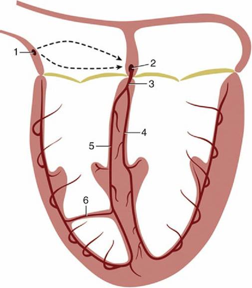
FIG. 7.15 Schematic drawing of the conducting system of the heart. The broken lines and arrows suggest the passage of the excitation wave through the atrial wall. 1, Sinoatrial node; 2, atrioventricular node; 3, atrioventricular bundle; 4, left limb; 5, right limb; 6, branch of right limb traversing the septomarginal band.
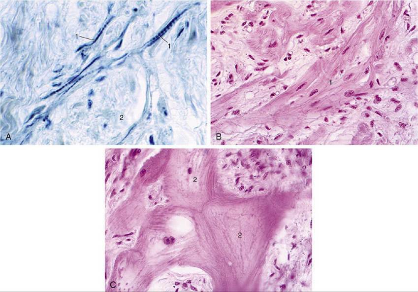
FIG. 7.16 (A) Sinoatrial node of the equine heart. 1, Nodal myofibers; 2, bundle of nerve fibers (hematoxylin and eosin [HE]; magnification ?279). (B) and (C) Atrioventricular node of equine heart (HE; magnification ?279). 1, Nodal myofibers; 2, Purkinje cells with abundant glycogen.