The Sympathetic System
The preganglionic axons of the sympathetic system originate from sympathetic preganglionic neurons located in the lateral gray column of the thoracolumbar part of the spinal cord (Fig.
8.73/1) and pass into the ventral roots of the thoracic and first several lumbar nerves. They continue into the spinal nerves and then leave the ventral rami, becoming the myelinated communicating branches (Fig. 8.73/2), which join the ganglia of the sympathetic trunk (Figs. 8.53/5 and 7 and 8.73/3). These bilateral trunks run the length of the neck and back, and each has a segmental arrangement, although strict correspondence of the ganglia with spinal nerves is evident only in the thoracic and cranial lumbar regions.The cervical part of the trunk begins at the large, spindle-shaped, cranial cervical ganglion placed close to the base of the skull (Fig. 8.73/5). The cervical trunk is associated with the vagus within the carotid sheath and forms the vagosympathetic trunk that proceeds down the neck. Whereas the vagus contains parasympathetic axons travelling caudally to innervate thoracic and abdominal viscera, the sympathetic trunk in the neck contains sympathetic axons travelling cranially to innervate structures of the head. The two components part company at the entrance to the chest, where the sympathetic trunk often bears a middle cervical ganglion by the first rib (Fig. 8.76/7'). The thoracic part of the sympathetic trunk then continues subpleurally, over the line of the costovertebral articulations, before passing dorsal to the diaphragm to enter the abdomen. Its thoracic part contains regularly spaced ganglia, although the first one or two are fused with caudal cervical ganglia to form the large cervicothoracic ganglion deep to the head of the first rib (Fig. 8.76/7). The lumbar part of the trunk, which lies between the psoas musculature and vertebral bodies, at first also carries a regular arrangement of ganglia, but the arrangement later becomes more erratic in that some caudal lumbar ganglia split into two or, less commonly, fuse with their neighbors.
The sacral part is even less regular, and right and left trunks may fuse, temporarily or finally, before extending into the tail, where it rapidly fades (Fig. 8.73/3).The locations of preganglionic sympathetic neurons are restricted to the thoracic and lumbar spinal segments, so it follows that only the thoracic and cranial lumbar ganglia are connected by myelinated communicating branches. However, all spinal and many cranial nerves are joined by unmyelinated communicating branches of postganglionic fibers destined for vessels, skin glands, and so forth. It should be stressed that the body wall and limbs are innervated only by these postganglionic sympathetic fibers. Sympathetic fibers contributing to most cervical nerves join within a single trunk, the vertebral nerve, which runs from the cervicothoracic ganglion through the foramina of successive cervical transverse processes (Fig. 8.73/7). The postganglionic sympathetic fibers traveling along with the first two cervical nerves and alongside cranial nerves extend from the cranial cervical ganglion; many form the internal carotid nerve that follows the internal carotid artery.
Several alternative fates are possibly with the preganglionic sympathetic fibers that enter the sympathetic chain, each to project on many ganglion cells. Some fibers synapse immediately within the local ganglion, others run cranially or caudally within the trunk to synapse within ganglia that are more cranial or caudal in the series, and yet others pass uninterruptedly through the trunk to proceed to a second set of (prevertebral) ganglia placed about the origin of the visceral branches of the abdominal aorta (Figs. 8.73/10 and 11 and 8.76/11 and 12). This last group constitutes the splanchnic nerves, which are rather variable in arrangement; usually one greater splanchnic nerve is formed by preganglionic fibers that leave the trunk from about the sixth to the penultimate thoracic ganglia, with lesser thoracic and lumbar splanchnic nerves arising at more caudal levels (Fig.
8.73/8 and 9).The viscera and vessels of the head receive their sympathetic innervation via the cranial cervical ganglion (Fig. 8.71/5). The postganglionic fibers that emerge from this ganglion radiate in a number of directions that carry them into the territories of the cranial nerves and the first two cervical nerves. Though many fibers pass through parasympathetic ganglia, they of course do so without interruption. The details are of rather limited clinical importance (although relevant to experimental work), and only a few points are presented here (see Fig. 8.71).
One large group of fibers follows the internal carotid artery into the cranial cavity and there provides twigs to the intracranial vessels and fiber bundles that join various nerves, especially the trigeminal and those to the extraocular muscles. Another group of fibers passes through the ciliary ganglion to the eyeball for ultimate distribution to the dilator pupillae. At a more proximal level, the internal carotid nerve gives off the deep petrosal nerve, which combines with the greater petrosal nerve (Fig. 8.71/11) in its passage through the pterygoid canal to the pterygopalatine ganglion (Fig. 8.71/7). These fibers are ultimately dispersed with the various nerves that supply structures within the orbit, nasal cavity, sinuses, and palate.
Other branches participate with parasympathetic fibers in forming a plexus within the tympanic cavity from which the parotid gland is supplied after passage beyond the otic ganglion. Yet other bundles of fibers entwine the external carotid artery and its branches.
The thoracic organs—heart, trachea, and lungs—are supplied by postganglionic fibers that form cardiac and pulmonary plexuses within the mediastinum after leaving the thoracic portion of the sympathetic trunk. These plexuses combine with the corresponding parasympathetic component (see Fig. 8.76).
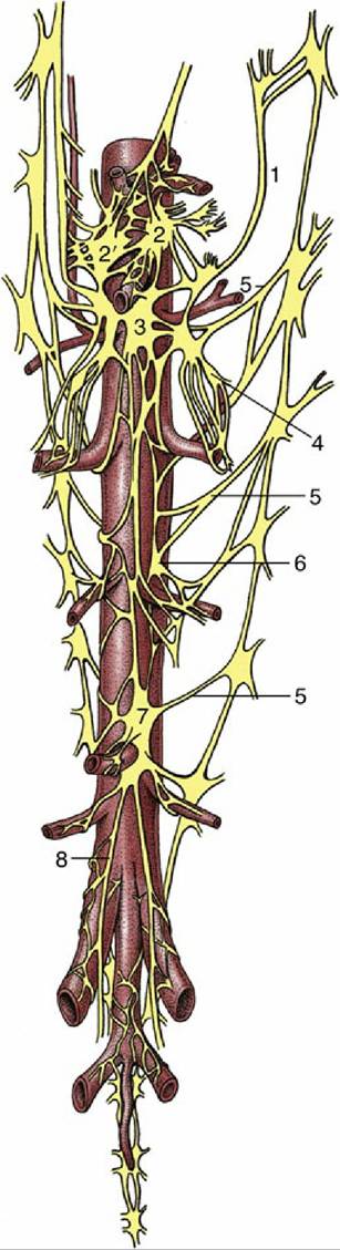
FIG.
8.77 Ganglia and plexuses of the abdominal cavity. Ventral view. 1, Greater splanchnic nerve ; 2, left celiac ganglion; 2', right celiac ganglion; 3, cranial mesenteric ganglion; 4, renal ganglion; 5, lumbar splanchnic nerves; 6, gonadal ganglion; 7, caudal mesenteric ganglion; 8, right hypogastric nerve.The abdominal and pelvic organs receive their sympathetic innervation through the various splanchnic nerves that lead to the celiac, cranial mesenteric, renal, aorticorenal, gonadal, and caudal mesenteric ganglia placed on the ventral face of the aorta by the origins of the visceral arteries. The preganglionic fibers synapse in these ganglia, and the postganglionic fibers that emerge from intricate plexuses (combining vagal contributions) enmesh, and run parallel to, the visceral arteries from which they obtain their names (Fig. 8.77).
» TABLE 8.3
Actions Controlled by the Autonomic Nervous System
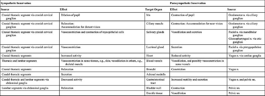
n., nerve; nn., nerves.
The pelvic organs are supplied with postganglionic fibers that leave the caudal mesenteric ganglion within the paired hypogastric nerves (Fig. 8.77/8). These fibres enter the pelvic cavity below the peritoneum to form a common pelvic plexus with the parasympathetic pelvic nerves (Fig. 8.76). As already mentioned, the sympathetic contribution to the pelvic plexus includes preganglionic sympathetic fibers that synapse in peripheral locations within the pelvis.