The Testes and Their Adnexa
The Testis
The testis combines endocrine and exocrine components within a common capsule. The endocrine component functions normally at the core temperature of the body, but in most mammals the successful production of the male gametes requires a temperature a few degrees lower than that within the abdomen.
However, spermatogenesis does occur normally at the core temperature in a few mammals (described as testicond, e.g., elephants, hyraxes) that have intra-abdominal testes. In many small mammals (chiefly found among rodents, insectivores, and bats) the testes descend from the abdomen into the scrotum transiently for the breeding season. This descent is brought about by contraction of the cremaster muscle sac found in these species.The testes are solid ellipsoidal organs whose bulk bears no fixed proportion to the body size. They are conspicuously small in cats and impressively large in sheep and goats. Their orientation also varies. They are carried with their long axes vertical in ruminants (necessitating a deep and pendulous scrotum), horizontal in horses and dogs, and tilted toward the anus in pigs and cats. These differences are broadly correlated with the position of the scrotum, which is below the caudal part of the abdomen in ruminants, perineal in pigs and cats, and intermediate in position in horses and dogs (Fig. 5.36). Each testis is separately suspended within the scrotum by a spermatic cord, a bundle of structures that includes the deferent duct and the supplying vessels and nerves enclosed within a double covering of peritoneum.
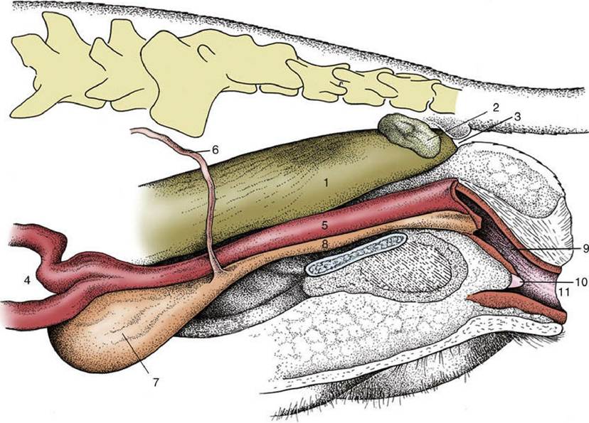
FIG. 5.35 Pelvic organs of the bitch. The lateral pelvic wall and the lateral wall of the vestibule have been removed. 1, Rectum; 2, anal sac; 3, anus; 4, uterus; 5, vagina; 6, ureter; 7, bladder; 8, urethra; 9, vestibule; 10, clitoris; 11, vulva.
The outer surface of the testis is made smooth by the direct peritoneal investment, except at the poles and along one margin, where the testis is attached to the epididymis, a structure formed by the coiled initial portion of the external duct system. The peritoneum covers a thickish capsule (tunica albuginea) composed mainly of dense connective tissue but sometimes including smooth muscle. The larger branches of the testicular artery and vein form a visible pattern within the capsule. The parenchyma is contained under moderate pressure and pouts through any incision of the capsule. Although slight swelling of the parenchyma can be accommodated by the assumption by the testis of a more globular form, any significant expansion raises the intratesticular pressure and produces severe pain, especially in inflamed testes (orchitis).* The capsule detaches septa and trabeculae that divide the parenchyma into lobules. The septa are not always conspicuous, but in those species in which they are well developed, they may be seen to converge on a substantial thickening (mediastinum testis), which may be axial or displaced toward the side bordering the epididymis (Fig. 5.37).
The soft, yellowish or brownish parenchyma consists of intermingled seminiferous tubules and interstitial tissue (Fig. 5.38). The greater part (60% in boars and stallions, 90% in rams and bulls) of the parenchyma is formed by the tubules, where spermatogenesis occurs. Each seminiferous tubule (Fig. 5.38) is much contorted and also looped so that both ends open into the rete testis (Fig. 5.38/5), a plexus of spaces within the mediastinum. Within the seminiferous tubules two cell types can be discerned: the Sertoli cells, which support and nourish the germ cells by the production of hormones and growth factors, and the seminiferous epithelium (Fig. 5.39). The rete drains by a dozen or so efferent ductules (Fig. 5.38/6) that pierce the capsule to join the head of the epididymis. The interstitial tissue consists of massed interstitial (Leydig) cells supported by a delicate connective tissue framework containing small blood and lymphatic vessels (Fig.
5.39).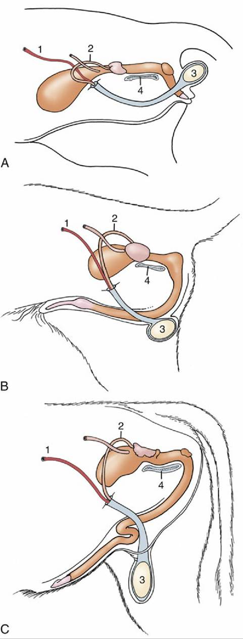
FIG. 5.36 The perineal, intermediate, and inguinal positions of the scrotum exhibited by the (A) tomcat, (B) dog, and (C) bull. 1, Testicular artery; 2, deferent duct; 3, testis; 4, pelvic symphysis.

FIG. 5.37 Median section of testis (bull). 1, Mediastinum testis; 2, testicular parenchyma.
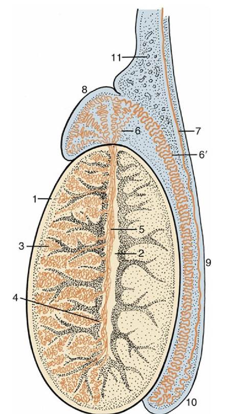
FIG. 5.38 Longitudinal section of a testis and epididymis, schematic. 1, Tunica albuginea; 2, mediastinum; 3, seminiferous tubules; 4, straight tubules; 5, rete testis; 6, efferent ductules; 6', epididymal duct; 7, deferent duct; 8, head of epididymis; 9, body of epididymis; 10, tail of epididymis; 11, pampiniform
plexus.
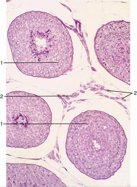
FIG. 5.39 Testis (dog) (140?). 1, Seminiferous tubules (showing spermatogenesis); 2, interstitial tissue with androgen-producing (Leydig) cells.
Endocrine Component
The endocrine functions of the testis are performed by the interstitial (Leydig) cells, responsible for androgen production, and the sustentacular (Sertoli) cells, responsible for inhibin production. Both types are normally under the pulsatile but more or less tonic control of gonadotropins (luteinizing hormone [LH] and follicle-stimulating hormone [FSH], respectively) produced in the pituitary (p. 204). Among other functions, the sustentacular cells produce activin and inhibin, which regulate the synthesis and release of FSH through mechanisms that may be direct or mediated via the hypothalamus. Androgens clearly have distinct local function but are also responsible for secondary sex characteristics such as the maturation of the accessory sex glands, male skeletomuscular development, skin characteristics, and even the prenatal differentiation of certain brain and spinal cord nuclei. These hormones are also partly responsible for the behavior typical of the male. They also act on hypothalamus to exert a negative feedback on pituitary gonadotropin secretion. In the fetal period, active production of androgens may take place without pituitary control. The interstitial cells in this period are also responsible for the production of insulin-like factor 3, which is associated with gubernacular outgrowth and thus with testicular descent. In the fetal period the sustentacular cells produce antimullerian hormone (AMH), which exerts an inhibitory effect on the paramesonephric ducts (p. 159), causing the
disappearance of most of the female duct system.
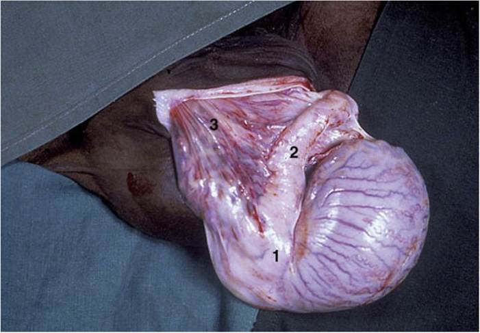
FIG. 5.40 Testis (horse). 1, Head of epididymis; 2, body of epididymis; 3, pampiniform plexus.