» The Thoracic Wall
Removal of the forelimbs exposes the contrasting form of the cranial and caudal parts of the thorax. The cranial part (formed by the sternal ribs) is narrow and bilaterally compressed and shows little movement.
The caudal part (formed by the asternal ribs) is conspicuously wider and more rounded and makes a substantial contribution to the respiratory excursions (see Fig. 20.8). In comparison with the bovine chest, the ribs are narrow and the intercostal spaces markedly wide, especially in their ventral parts. The arrangement of the structures within the spaces follows the usual pattern.The short, stout first rib is almost immobile because it is stabilized by tight joints with the vertebral column and sternum and by anchorage to the cervical vertebrae through the scalenus muscle. The brachial plexus divides this muscle into ventral and (small) middle parts, while the axillary vessels emerge ventral to it. These vessels wind around the cranial margin of the first rib, where the axillary artery may be palpated against the bone. Previously, the artery was punctured at this site when a sample of arterial blood was required (Fig. 20.3/1'), but currently the carotid artery is preferred.
In conformity with the length of the thorax, the diaphragm is more oblique than in other domestic species but has the same general form. It bulges forward from its peripheral attachments to the lumbar vertebrae, ribs, and sternum. Its most cranial part, the vertex, is situated directly above the sternum and projects on the lower part of the sixth space or preceding rib. The dorsal part of the diaphragm is molded to present right and left elevations between which the median portion is retracted by the crura to form a recess. The middle and ventral parts are uniformly curved from side to side. The openings within the diaphragm show no important specific features (Fig.
20.4).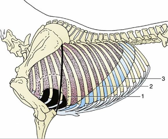
FIG. 20.1 Projections of the heart and lung on the left thoracic wall. The heavy line indicates the caudal border of the triceps. 1, Outline of heart; 2, basal border of lung; 3, line of pleural reflection.
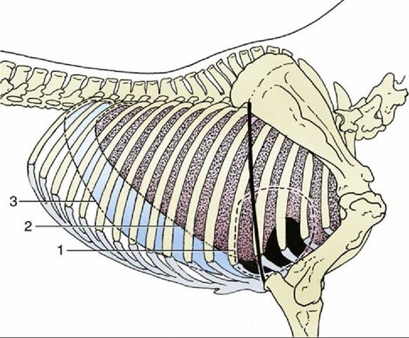
FIG. 20.2 Projections of the heart and lung on the right thoracic wall. The heavy line indicates the caudal border of the triceps. 1, Outline of heart; 2, basal border of lung; 3, line of pleural reflection.
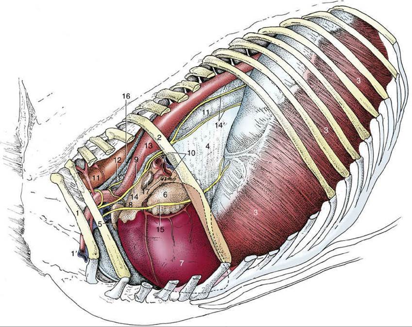
FIG. 20.3 Structures within the mediastinum. The mediastinal pleura cranial to the heart has been removed, which exposes the cranial lobe of the right lung. 1, First rib; 1', axillary vessels; 2, sixth rib; 3, diaphragm; 4, caudal mediastinum covering right lung; 5, right auricle; 6, left auricle; 7, left ventricle; 8, pulmonary trunk; 9, ligamentum arteriosum; 10, root of lung; 11, esophagus; 12, trachea; 13, aorta; 14, vagus nerve; 14', dorsal and ventral vagal trunks; 15, phrenic nerve; 16, thoracic duct; 17, tracheobronchial lymph nodes.
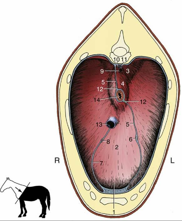
FIG. 20.4 Cranial surface of the diaphragm. 1, Sternal and costal parts of diaphragm; 2, tendinous center; 3, left crus; 4, right crus; 5, caudal mediastinum; 6, left phrenic nerve; 7, plica venae cavae; 8, right phrenic nerve; 9, aorta; 10, right azygous vein; 11, thoracic duct; 12, dorsal and ventral vagal trunks; 13, caudal vena cava; 14, esophagus; L, left side; R, right side.