THE THORACIC WALL AND PLEURA (See also pp. 41-43, 48-52, and 158-160.)
The dog generally has 13 rib pairs of which nine are sternal. Asymmetry of number and the presence of 12 or 14 pairs are both occasionally found. The first three to four ribs are almost vertical; behind this, the ribs slope increasingly caudoventrally (see Figure 2-1).
The ribs are relatively narrow, resulting in wide intercostal spaces, which is an advantage in thoracic surgery. The costal cartilages at first continue the direction of the bony ribs but then bend forward, almost at right angles (see Figure 13-6), to form the rib “knees.” Those of the sternal ribs form synovial articulations with the sternum, which allow expansion of the thorax when the ribs are carried cranially in the “bucket-handle” movement. The cartilages of the four asternal ribs join to form the costal arch, which is easily palpated and may be followed to the vicinity of the xyphoid cartilage (Figure 13-8/5). The slender, cylindrical sternebrae are slightly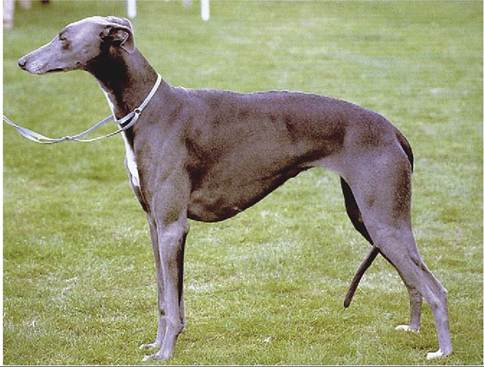
Figure 13-1 Deep and laterally compressed thorax of the Greyhound.
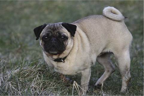
Figure 13-2 Broad, barrel-shaped thorax of the Pug.
thickened at their extremities where the costal cartilages attach. Only a thin layer of compact bone encloses the spongy interior, and this, combined with the superficial position, makes them ideal for bone marrow biopsy.
The intercostal spaces have the usual construction. The principal intercostal vessels and nerves run caudo- medially to the ribs, under the endothoracic fascia. Additional vessels from the internal thoracic trunks follow the cranial borders of the ribs in the ventral parts of the spaces (see Figure 13-4). These locations must be borne in mind when contemplating incision or puncture.
When such procedures are contemplated, a useful guide to the topography is supplied by the boundary between the scalenus and external abdominal oblique muscles, which marks the fifth intercostal space. The space chosen for a lateral thoracotomy is not always that suggested by prior knowledge of the topography or by preliminary radiography; the ribs are so much more easily displaced cranially than caudally that a more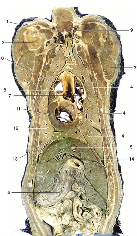
Figure 13-3 Dorsal section of the canine trunk level with the base of the heart, dorsal view. 1, Cephalic vein; 2, proximal end of humerus; 3, triceps; 4, cranial, middle, caudal, and accessory lobes of the right lung; 5, liver; 6, stomach; 7, right atrium; 8, aortic arch; 9, cranial vena cava; 10, pulmonary valve; 11, left atrioventricular valve; 12, divided cranial and caudal lobes of the left lung; 13, caudal mediastinum; 14, diaphragm.
favorable exposure of the “target” region may be gained by opening the space immediately caudal to the one that initially seemed most appropriate.
The diaphragm arises by right and left crura from the first few lumbar vertebrae and attaches to the medial surfaces of the ribs close to the costal arches and to the sternum. Its strong curvature brings its most cranial point to the level of the sixth or seventh rib. The small, triangular tendinous center transmits the caudal vena cava a little to the right of the median plane. The openings for the esophagus and aorta lie in the fleshy lumbar
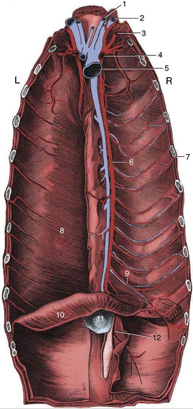
Figure 13-4 The vessels on the floor of the canine thorax; the transversus thoracis has been removed on the right. 1, Internal jugular vein; 2, external jugular vein; 3, vertebral artery; 4, right subclavian artery; 5, cranial vena cava; 6, internal thoracic artery; 7, intercostal artery; 8, transversus thoracis; 9, musculophrenic artery; 10, diaphragm; 11, xiphoid cartilage; 12, cranial epigastric artery.
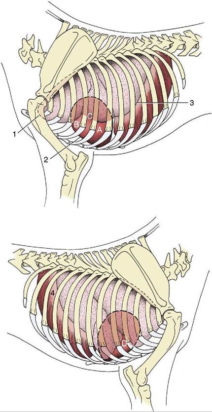
Figure 13-5 Left and right surface projections of the canine heart and lungs. Circled letters on the heart: puncta maxima of left atrioventricular valve (A), pulmonary valve (B), aortic valve (C), and right atrioventricular valve (D). 1, Apex of left lung (broken line) in cupula pleurae; 2, heart; 3, basal border of lung; 4, diaphragm.
part, and the former is opposite the upper palpable part of the tenth rib (Figure 13-9). In lateral radiographs the strongly convex ventral part of the diaphragm presents a simple border that is continued dorsally by the paired outlines of the cupulae (Figure 13-10, A/4); the more cranial outline of this double image is provided by the cupula on the “lower” side of a laterally recumbent animal, which is the side subjected to greater forward pressure from the abdominal viscera. A further guide to the correct identification of the twin elevations is provided by the gas bubble that is usually found in the gastric fundus, which is of course located on the left side. The doubling of the outline is less distinct in cats in which the lighter abdominal organs exert possibly less pressure.
A sudden increase in abdominal pressure, commonly produced by compression in traffic accidents, may tear the diaphragm and allow abdominal viscera to enter the thoracic cavity (diaphragmatic hernia).
At rest, ventilation principally depends on the diaphragm, but when respiratory demands increase, other muscles are called into play. Some or all of the
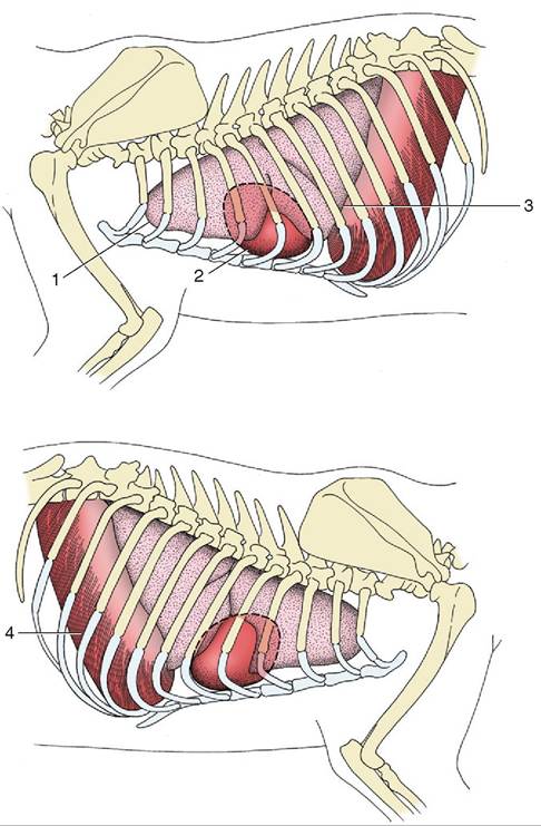
Figure 13-6 Left and right surface projections of the feline heart and lung. 1, Apex of left lung; 2, heart; 3, basal border of lung; 4, diaphragm.
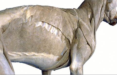
Figure 13-7 Notice the attachment of the external abdominal oblique muscle on the ribs.
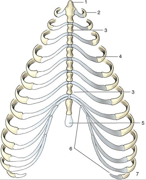
Figure 13-8 Canine sternum and costal cartilages, ventral view. 1, Manubrium; 2, first rib; 3, sternebra; 4, costochondral junction; 5, xiphoid cartilage; 6, costal arch; 7, floating rib.
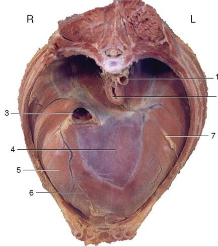
Figure 13-9 Cranial view of the canine diaphragm. 1, Aorta; 2, esophagus; 3, caudal vena cava; 4, tendinous center; 5, sternal and costal parts of diaphragm; 6, attachment of plica venae cavae; 7, attachment of caudal mediastinum.

Figure 13-10 Lateral (A) and ventrodorsal (B) bronchograms of the right canine lung. 1, Sternum; 2, heart; 3, liver behind diaphragm; 4, paired shadows of the cranial extent of the diaphragm; 5, scapula.
external intercostal, Sternocephalic, ventral serratus, and scalenus may be used to assist at inspiration, whereas the internal intercostal and abdominal muscles may assist at expiration.
The pleural cavities present the usual features, of which the most important clinically are the cupulae cranially, the caudal reflection of the costal pleura onto the diaphragm, and the presence and extents of the costomediastinal and costodiaphragmatic recesses. In the dog, the cupulae (see Figure 13-5) project only slightly in front of the first ribs, but this is sufficient to make it possible for air to be introduced into a pleural cavity by a penetrating wound that appears to be confined to the base of the neck, the result of which is the collapse of a lung.
The junction between costal and diaphragmatic pleura, the line of pleural reflection, defines the caudal extent of the pleural cavity. The line runs from the sternum along the eighth costal cartilage, crosses the middle of the ninth cartilage, and then proceeds in a curve that intersects the eleventh costochondral junction to reach the dorsal end of the last rib. The two recesses are of course never fully exploited by the lungs. Fluid may be collected through the ventral third of any of the fourth to seventh intercostal spaces of a dog standing or restrained in sternal recumbency. In cases of pneumothorax, air may be aspirated at the dorsal part of the seventh or eighth space of dogs similarly placed. The eighth space is optimal for this purpose in the cat.
The coupling of the lung to the thoracic wall, maintained by a thin layer of pleural fluid, is disrupted when air gains entry to the pleural cavity. This causes not only collapse of the lung but also expansion of the rib cage as the thoracic wall recoils outward. Although pneumothorax is generally produced by trauma of the thoracic wall, it may result from rupture of the lung or trachea or from perforation of the esophagus.