THE THYROID GLAND
The thyroid gland lies on the trachea directly behind, and sometimes overlapping, the larynx. Its form varies greatly: in the dog and the cat the gland consists of separate masses that are occasionally connected by an isthmus (Figure 6-4, A); in the horse, paired lobes are widely dissociated but connected by an insubstantial isthmus (Figure 6-4, B); in cattle the lobes are connected by a wide isthmus of parenchymal tissue (Figure 6-4, C); in small ruminants the isthmus is inconstant
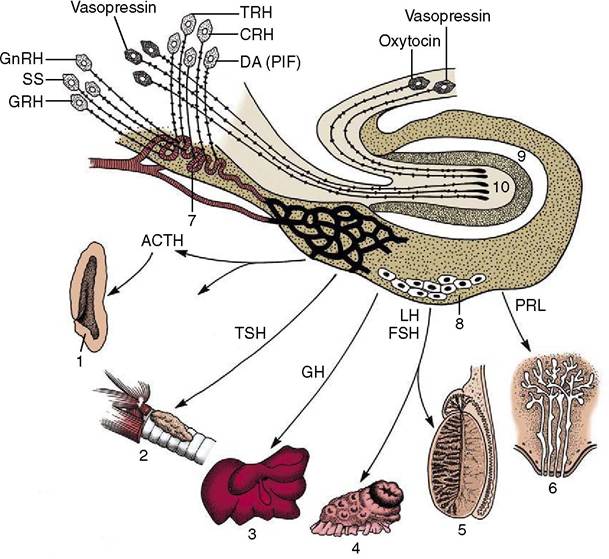
Figure 6-3 Organization of the brain-pituitary-peripheral organ axis.
TRH, thyrotropin-releasing hormone; CRH, corticotropinreleasing hormone; DA, dopamine; PIF, prolactin-inhibiting factor; GnRH, gonadotropin-releasing hormone; SS, somatostatin; GRH, growth hormone-releasing hormone; ACTH, adrenocorticotropic hormone; TSH, thyroid-stimulating hormone; GH, growth hormone; LH, luteinizing hormone; FSH, follicle-stimulating hormone; PRL, prolactin. 1, Adrenal cortex; 2, thyroid; 3, liver; 4, ovary; 5, testis; 6, mammary gland; 7, median eminence; 8, anterior lobe of pituitary; 9, intermediate lobe of pituitary; 10, neural lobe of pituitary.and when present is a mere connective tissue strand. In yet other species the thyroid has a more compact form and exhibits a relatively large median (pyramidal) lobe in addition to the lateral lobes. This arrangement, found in pigs and human subjects, provides a cover on the trachea that extends toward the thoracic inlet (Figure 6-4, D); it explains the name given to the gland.
The gland has its origin in a median outgrowth from the part of the pharyngeal floor that contributes to the tongue (p. 142). The primordium extends caudally on the ventral surface of the trachea before dividing at its apex into divergent processes that extend dorsolaterally to reach the boundary between the trachea and the esophagus (Figure 6-5/2).
In most mammals the connection with the developing tongue (thyroglossal duct) is never patent and it later regresses in its entirety.The mature gland is enclosed within a connective tissue capsule that is loosely attached to neighboring organs. Its substance, generally brick-red, obtains a rather granular texture from the many enclosed follicles of which it is composed. In some species (e.g., cattle) these give the intact organ an irregular appearance, but in others (e.g., dog) the surface is quite smooth. The tissue is relatively firm, and this consistency, allied to the form, size, and location, enables the lobes to be identified in larger species by palpation caudal to the larynx. They are not palpable in the healthy dog.
The size of the thyroid gland varies greatly, depending to a large extent on the iodine content of the diet; when this content is deficient, enlargement (goiter) may develop, and in some parts of the world it is customary to add iodine to table salt as a preventive measure. In dogs the relative weight of the thyroid may vary by a factor of as much as six, although the increasing use of commercial foods (of uniform composition) now tends to reduce this variation. Average dimensions in mediumsized dogs are of the order of 6 ? 1.5 ? 0.5 cm. Accessory masses of thyroid tissue are sometimes located along the cervical trachea and are occasionally carried into the thorax by the descending heart.
The gland is mainly supplied by the cranial thyroid artery, which arises from the common carotid artery and arches around the cranial pole. A subsidiary supply is occasionally provided by a caudal thyroid artery,
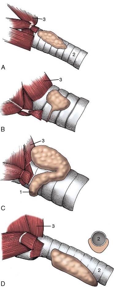
Figure 6-4 The thyroid gland of the dog (A), horse (B), cattle (C), and pig (D). The inset to D illustrates the subtra- cheal connection in transverse section in the pig. 1, Isthmus; 2, trachea; 3, cricopharyngeus.
which takes a more proximal origin.
In the dog the two vessels are connected by a substantial anastomosis along the dorsal margin. The venous drainage is to the internal jugular vein. The glandular tissue receives both sympathetic and parasympathetic fibers; the former is routed through the cranial cervical ganglia, the latter through the laryngeal branches of the vagus nerves. The fibers are predominantly vasomotor, and denervation has little effect on secretory activity.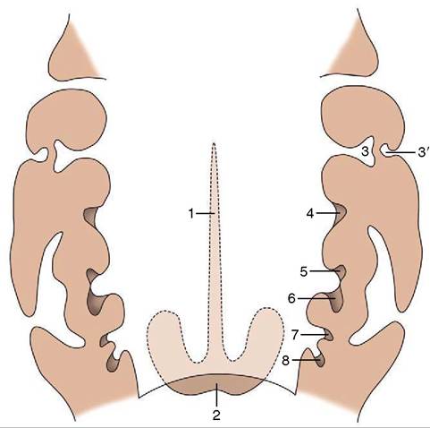
Figure 6-5 The pharyngeal primordia of certain endocrine structures; dorsal view, schematic. 1, Thyroglossal duct; 2, thyroid gland; 3, first pharyngeal pouch; 3', external acoustic meatus; 4, palatine tonsil (second pouch); 5, parathyroid III; 6, thymus; 7, parathyroid IV; 8, ultimobranchial body.
The main lymph drainage of the thyroid in the dog proceeds to the cranial deep cervical nodes.
The thyroid hormones, concerned with metabolism and growth, are produced by the follicular cells that compose the bulk of the parenchyma. They are stored in the follicular fluid and later broken down to yield the final products, which are released into the bloodstream.
A small portion of the parenchyma is provided by parafollicular (or C) cells. These appear to have their origin in the ultimobranchial bodies that derive from epithelial clusters of the fourth pharyngeal pouches that are invaded by neural crest cells (Figure 6-5/8). C cells produce calcitonin, a hormone antagonistic to parathormone in some species. It also seems to play a role in fetal bone growth, and it protects the maternal skeleton against excessive demineralization.