The Trigeminal Nerve (V)
The trigeminal nerve, the largest of the cranial nerves, is sensory to the skin and deeper tissues of the face and provides motor innervation to the muscles of first pharyngeal (mandibular) arch origin.
Proprioceptive afferent fibers, which include many from muscles that receive their motor innervation from other cranial nerves, pass to the rostral trigeminal mesencephalic nucleus; the other exteroceptive afferent fibers synapse onto the pontine and spinal trigeminal nuclei (Fig. 8.25/7). The efferent fibers originate in the trigeminal motor nucleus (Fig. 8.25/17). The peripheral nerve itself is formed by the fusion of sensory and motor roots that attach to the ventrolateral aspect of the pons. The larger sensory root carries the massive trigeminal ganglion and, just beyond this, divides into the three primary branches (ophthalmic, maxillary, and mandibular) that give the trunk its name. The mandibular branch unites with the motor root to constitute the mixed mandibular nerve; the ophthalmic and maxillary divisions remain purely sensory at this level, although peripheral connections with other cranial nerves introduce somatic and visceral efferent fibers into certain branches. The mandibular nerve emerges through the oval foramen in the floor of the cranial cavity. The ophthalmic and maxillary nerves run rostrally to emerge through the orbital fissure and round foramen, respectively (in ruminants the two openings are combined).The three primary divisions are initially each restricted to a different process of the embryonic face, a fact that explains the crisply defined adult territories (cf. the dermatomes of the trunk). The ophthalmic nerve supplies the frontonasal process, the primordium of the forehead and nose regions; the maxillary nerve supplies the maxillary process, the primordium of the upper jaw, and associated parts; and the mandibular nerve supplies the mandibular process, the primordium of the lower jaw, and associated parts, which include the masticatory and other first pharyngeal arch muscles (Fig.
8.69).The ophthalmic nerve (Fig. 8.69/1), for which the convenient notation is V-1, divides into three divergent branches (lacrimal, frontal, nasociliary) soon after entering the orbit. The lacrimal nerve (Fig. 8.69/3) passes to the lateral part of the orbital perimeter and, after detaching branches to the lacrimal gland and other deeper structures, emerges to supply the skin about the lateral angle of the eye. The more considerable territory of the frontal nerve (Fig. 8.69/2) includes much of the upper eyelid, the forehead, and, through branches that penetrate the bone, the mucosa of the frontal sinus.
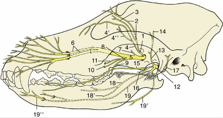
FIG. 8.69 Distribution pattern of the trigeminal nerve (n.) of the dog. 1, Ophthalmic n.; 2, frontal n.; 3, lacrimal n.; 4, nasociliary n.; 4', infratrochlear n.; 4", long ciliary n.; 5, maxillary n; 6, infraorbital n.; 7, zygomatic n.; 8, pterygopalatine n.; 9, lesser palatine n.; 10, greater palatine n.; 11, caudal nasal n.; 12, mandibular n.; 13, masticatory n.; 14, deep temporal n.; 15, buccal n.; 16, pterygoid n.; 17, auriculotemporal n.; 18, lingual n.; 18', sublingual n.; 19, inferior alveolar n.; 19', mylohyoid n.; 19", mental n.
The nasociliary nerve (Fig. 8.69/4) runs toward the medial wall of the orbit. One branch, the infratrochlear nerve (Fig. 8.69/4'), emerges on the face after supplying structures at the medial angle; it supplies another portion of the mucosa of the frontal sinus and in small ruminants detaches the principal nerve to the horn. Other branches of the nasociliary nerve include long ciliary and ethmoidal nerves. The long ciliary nerves (Fig. 8.69/4') penetrate the posterior aspect of the eyeball to supply sensitive tissues, including the cornea; the ethmoidal nerve first reenters the cranial cavity through the ethmoidal foramen and subsequently passes to the nasal cavity via the cribriform plate before dividing into medial and lateral branches to the mucosa.
The maxillary nerve (V-2) runs across the wall of the pterygopalatine fossa ventral to the orbit (Fig. 8.69/5). It bears, or lies close to, the pterygopalatine ganglion, but the relationship is purely topographic. It then enters the infraorbital canal at the maxillary foramen, where it becomes known as the infraorbital nerve (Fig. 8.69/6) in anticipation of its reappearance on the face at the infraorbital foramen.
Collateral branches of the maxillary nerve detach within the pterygopalatine fossa. They include the zygomatic nerve (Fig. 8.69/7), which supplies the lower eyelid and adjacent skin and is the origin of the principal nerve of the horn in cattle.
The second branch of the maxillary nerve, the pterygopalatine nerve (Fig. 8.69/8), detaches the lesser palatine nerve (Fig. 8.69/9) to the soft palate; the greater palatine nerve (Fig. 8.69/10), which reaches the hard palate after traversing the palatine canal and supplies both the palatine mucosa and the floor of the nasal vestibule; and the caudal nasal nerve (Fig. 8.69/11), which passes through the pterygopalatine foramen to supply mucosa of the ventral part of the nasal cavity, maxillary sinus, and palate.
Within the infraorbital canal, the infraorbital nerve (Fig. 8.69/6), being the continuation of the maxillary nerve, detaches short twigs to the alveoli of the cheek teeth and nasal mucosa and longer rostral alveolar branches that continue within the bone, beyond the infraorbital foramen, to the alveoli of the canine and incisor teeth. After emerging at the infraorbital foramen, the infraorbital nerve supplies various labial and nasal branches to the structures of the muzzle, including some branches that run back over the nose to the edge of the infratrochlear territory. Although covered by muscle at its emergence from the infraorbital foramen, the infraorbital nerve can usually be palpated, stimulated by pressure, or blocked by injection of local anesthetic solution.
On leaving the cranium, the mandibular nerve (V-3) detaches several branches in close succession that pass to the masseter, temporalis, medial and lateral pterygoid, tensor veli palatini, and tensor tympani muscles (Fig.
8.69/12). There are minor variations in their pattern, and the nerves to the masseter and temporalis are often initially joined as a short masticatory nerve (Fig. 8.69/13). The masseteric nerve passes to the masseter muscle between the coronoid and condylar processes of the mandible. The deep temporal nerves (Fig. 8.69/14) run dorsomedially to the temporalis muscle. The otic ganglion lies close to the origin of the pterygoid nerves (Fig. 8.69/16).The next branch from the mandibular nerve, the buccal nerve (Fig. 8.69/15), is sensory to the tissues of the cheek, which it reaches after first passing between the pterygoideus and temporalis and then between the maxillary tuber and mandible. Its origin is followed by that of the auriculotemporal nerve (Fig. 8.69/17), which bends around the caudal border of the mandible to enter the face a little ventral to the temporomandibular joint. It is sensory to the skin of the temporal region and over much of the external ear, including the lining of the canal leading to the eardrum. It continues onto the face as the transverse facial branch, supplying a strip of skin extending to the corner of the mouth.
The mandibular nerve continues between the medial and lateral pterygoid muscles before dividing into its end-branches, the lingual and inferior alveolar nerves.
The lingual nerve (Fig. 8.69/18) detaches twigs to the oropharyngeal mucosa before dividing into a deep branch that enters the tongue, and a superficial branch, the sublingual nerve (Fig. 8.69/18'), that runs medial to the mylohyoideus below the mucosa of the oral floor that it supplies. The branch of the lingual nerve to the tongue is joined by the chorda tympani, a branch of the facial, which contains axons of preganglionic visceral efferent fibers to salivary glands that synapse in the adjacent mandibular ganglion as well as gustatory (special visceral afferent) fibers innervating the taste buds of the rostral two-thirds of the tongue. Other sensory fibers in the lingual nerve supply general sensation (general somatic afferent) in the same rostral two-thirds of the lingual mucosa.
The inferior alveolar nerve (Fig. 8.69/19) detaches the mylohyoid nerve (Fig. 8.69/19') to supply motor innervation to the mylohyoideus and rostral belly of the digastricus before entering the mandibular canal at the mandibular foramen. The inferior alveolar nerve supplies sensory innervation to the lower cheek teeth before a large part reappears at the mental foramen as the mental nerve (Fig. 8.69/19", which supplies tissues of the lower lip and chin. In some species several mental branches exit through as many foramina. Although also covered by muscle, the mental nerve(s) can be palpated, compressed, and blocked at their emergence from the foramina.
Injuries to, or disease of, the branches of the trigeminal nerve produce sensory deficiencies in their territories and sometimes manifest as chronic facial irritation; some branches are frequently blocked for minor surgery of the head. Destructive lesions of the mandibular nerve produce paralysis of the muscles that close the jaw; when the lesion is unilateral, the resulting atrophy may be more obvious than any motor disability. A temporary idiopathic bilateral paralysis of the trigeminal musculature, characterized by a dropped jaw, has been reported in dogs.