THE TROCHLEAR NERVE (IV)
The trochlear nerve, which is small, is motor to the dorsal oblique muscle. The nucleus of origin within the tegmentum of the midbrain gives rise to a fiber bundle that decussates internally before emerging from the rostral medullary velum (Figure 8-23/nerve include long ciliary and ethmoidal nerves.
The long ciliary nerves (Figure 8-68/4") penetrate the posterior aspect of the eyeball to supply sensitive tissues, including the cornea; the ethmoidal nerve first re-enters the cranial cavity through the ethmoidal foramen and subsequently passes to the nasal cavity via the cribriform plate before dividing into medial and lateral branches to the mucosa.The maxillary nerve (V-2) runs across the wall of the pterygopalatine fossa ventral to the orbit (Figure 8-68/5). It bears, or lies close to, the pterygopalatine ganglion, but the relationship is purely topographical. It then enters the infraorbital canal at the maxillary foramen, where it becomes known as the infraorbital nerve (Figure 8-68/b) in anticipation of its reappearance on the face at the infraorbital foramen.
Collateral branches detached within the pterygopalatine fossa include the zygomatic nerve (Figure 8-68/7), which supplies the lower eyelid and adjacent skin and is the origin of the principal nerve of the horn in cattle.
The second branch, the pterygopalatine nerve (Figure 8-68/nucleus (III); 2, parasympathetic facial nucleus (VII); 3, parasympathetic glossopharyngeal nucleus; 4, parasympathetic vagus nucleus; 5, cranial cervical ganglion; 6, ciliary ganglion; 7, pterygopalatine ganglion; 8, mandibular ganglion; 9, otic ganglion; 10, short ciliary nerves; 11, greater petrosal nerve; 12, deep petrosal nerve; 13, chorda tympani; 14, tympanic plexus, short petrosal nerve.
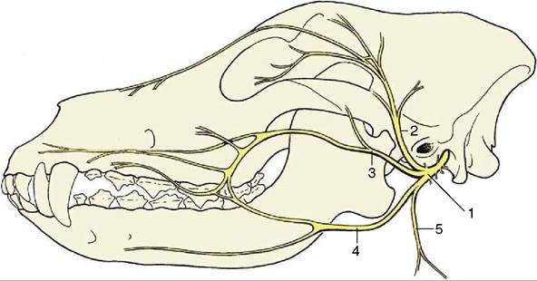
Figure 8-69 Distribution pattern of the facial nerve of the dog.
1, Facial n.; 2, auriculopalpebral n.; 3, dorsal buccal branch; 4, ventral buccal branch; 5, cervical branch.the middle ear. The next branch, the chorda tympani (Figure 8-70/13), crosses the tympanic cavity to emerge at the petrotympanic fissure, after which it converges on and becomes incorporated within the lingual branch of the mandibular nerve (p. 353).
The first branches of the free portion of the facial nerve are the internal and caudal auricular nerves, which supply muscles of the external ear and other branches to some hyoid muscles, including the caudal belly of the digastricus. The main trunk enters the face by turning around the mandible, where it is first contained between the masseter and the parotid gland. It divides at about this level (although there are species differences) into three terminal branches.
In some species the auriculopalpebral nerve (Figure 8-69/2) is detached before the main trunk reaches the face, and it is then less vulnerable to injury from superficial trauma to the side of the head. It crosses the zygomatic arch, heading for the space between the upper eyelid and external ear, before dividing into branches that supply the muscles of the eyelids (excluding levator palpebrae superioris) and the auricular muscles in front of the external ear.
The dorsal buccal branch (Figure 8-69/3), which may take the form of a leash of divergent branches, crosses the masseter en route to the muzzle.
In some species the ventral buccal branch (Figure 8-69/4) may take a similar path at a slightly more ventral level, but in others it takes a divergent course, first running within the intermandibular space before entering the face with the parotid duct and facial vessels, where they cross the mandible in front of the masseter. Together, the buccal branches supply the muscles of the cheek, lips, and nostrils. Their peripheral branches join with those of the trigeminal nerve at various levels, and many of the smaller trunks combine motor (facial) and sensory (trigeminal) fibers.
The effects of injury or disease clearly depend on the site of the lesion. Lesions that are situated more centrally, which tend to have more sinister origins, affect the whole facial field and lead to loss of secretory activity by the lacrimal and salivary (except the parotid) glands in addition to muscular paralysis. Lesions involving the main trunk near its exit from the bone paralyze the entire mimetic musculature, while more peripheral lesions may spare some groups, depending on their site and specific and individual variations in the branching pattern. Those confined to the auriculopalpebral nerve produce drooping of the external ear and narrowing of the palpebral fissure with inability to close the eye. Damage to the buccal branches may paralyze the muscles of the lips and cheeks, allowing a quid of food to collect in the oral vestibule. It may also lead to deformation of the muzzle, which is drawn out of symmetry by the unopposed activity of the muscles on the sound side. The alteration in appearance is not always very striking, and the uninjured side, to which the muzzle is drawn, may sometimes appear to have the more distorted aspect. The distortion tends to be more pronounced in the horse and sheep than in other domestic species. (It is important to be aware that in unilateral facial spasm, seen occasionally in the dog, the nose may be drawn toward the affected side.)
The auriculopalpebral nerve is sometimes blocked to facilitate examination of the eye.