THE UDDER
The four mammary glands of the cow are consolidated in a single mass, the udder, placed below the caudal part of the abdomen and extending between the thighs. The udder is divided into quarters corresponding to the four glands, and each bears a principal teat.
A median groove divides the udder into right and left halves, but the boundary between a forequarter and a hindquarter is rarely distinct. Most of the dorsal base is shaped to fit against the belly wall, but the part below the pelvis is narrower because it is compressed between the thighs (Figure 29-38). The skin over the udder is thin, supple, and mobile, except over the teats, where it is tightly bound down and naked.The udder is suspended by strong sheets of fascia that surround and enclose the gland substance and are continuous with the connective tissue framework that permeates the entire organ. The fascia forms a continuous investment over each half, but it is customary to describe medial and lateral laminae as though these were independent formations. The medial lamina arises mainly from the tunica flava, in small part from the symphysial tendon, and is largely composed of elastic tissue. The lateral lamina arises from the external crus of the inguinal ring and, behind this, from the medial femoral fascia and is composed of dense connective tissue (Figures 29-39 and 29-40). Both laminae thin when followed ventrally, which is the result of their detachment of numerous leaves that interdigitate with layers of glandular tissue. The different natures of the two laminae explain the sagging of the medial part of the heavily laden udder. Ever increasing demands for milk production place a heavy and sometimes unsustainable burden on the suspensory apparatus, which occasionally ruptures—a disastrous happening.
Each gland is constructed about a branching duct system, separated from its neighbors by connective tissue.
The alveolar secretory units lead to small excretory ducts that combine with others until, after several successive unions, about a dozen wide lactiferous ducts are produced; these converge on a large sinus situated in the lower part of the quarter and extend into the teat (Figure 29-41). The lactiferous ducts are unusual in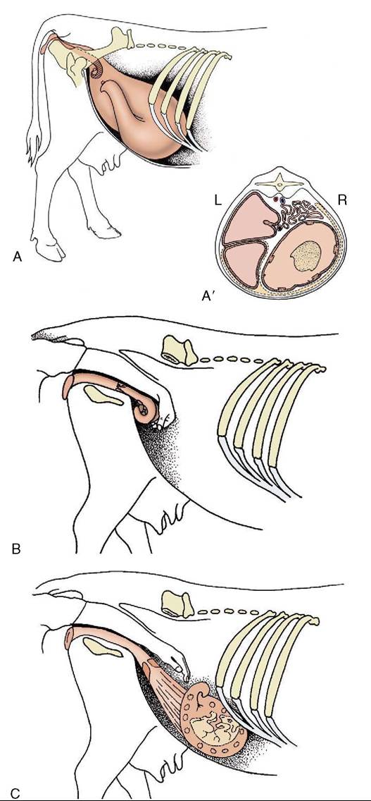
Figure 29-37 The position of the nongravid uterus and various stages of the gravid uterus in lateral view. A, Nongravid and 6-months gravid uterus. A', The topography of the 6-months gravid uterus in transverse section. B, At 2 to 3 months the uterus has begun to slide down the caudal abdominal wall, but it can be scooped up by the hand in the colon. C, At 5 months the uterus is temporarily out of reach.

Figure 29-38 Holstein cow with well-developed udder.
1, Mammary vein.
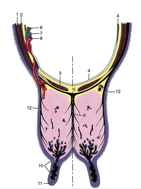
Figure 29-39 Transverse section of the abdominal floor and cranial quarters of the bovine udder. 1, External abdominal oblique; 2, internal abdominal oblique; 3, rectus abdominis; 4, peritoneum; 5, linea alba; 6, lymph vessel; 7, external pudendal vein; 8, external pudendal (mammary) artery; 9, medial laminae of suspensory apparatus; 10, lactiferous sinus; 11, papillary duct; 12, lateral laminae of suspensory apparatus.
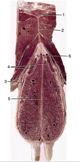
Figure 29-40 Transverse section of the pelvic floor and caudal quarters of the bovine udder. 1, Pelvic symphysis; 2, symphysial tendon; 3, lateral suspensory laminae; 4, mammary (superficial inguinal) lymph node; 5, medial suspensory laminae; 6, tributary of external pudendal vein.
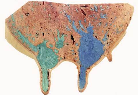
Figure 29-41 Sagittal section of udder, showing teat and gland sinuses and lactiferous ducts filled with latex (cranial quarter [green]; caudal quarter [blue])
demonstrating alternating wider and narrower sections.
The more superficial dilations, which may be 3 cm or more in caliber, may be palpable when distended with milk and are then known as “milk knots.” Although the duct systems are independent, infection readily spreads between the quarters of the same side.The lactiferous sinus has a capacity of several hundred milliliters and is divided by a mucosal fold into gland and teat parts. The fold, based on a submucosal ring of veins, varies in prominence; occasionally it may be sufficiently pronounced to impede milk flow.
The teats, though variable, are most often cylindrical and about 8 cm long. The teat wall, generally about 6 mm thick, increases to about 1 cm at the lower end, where it is traversed by the papillary duct (Figure 29-42). The wall consists of a dry, outer skin, an intermediate layer that includes smooth muscle and many veins and constitutes a form of erectile tissue, and an inner mucosal layer marked by folds. The lining, generally yellowish, is white in the papillary duct, where it shows a pattern of low ridges; these, when followed proximally, are found to radiate from the upper opening, although it must be admitted that the arrangement is rarely as distinct as traditionally described (Figure 29-43). Desquamation of the epithelium provides a bacteriostatic substance that helps occlude the passage. A more effective means of closure is provided by a sphincter muscle, reinforced by elastic tissue.
Accessory teats, sometimes associated with functional glandular tissue, are very common. They are undesirable as they may be a complication at milking.
The vascular arrangements are necessarily generous. The main supply, which continues the external pudendal artery, has a diameter that may exceed 15 mm where it passes through the inguinal canal accompanied by a satellite vein, lymphatics, and nerves (see Figure 29-39). On reaching the base of the udder, it divides into divergent branches, one passing cranially, the other caudally;
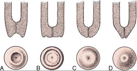
Figure 29-42 Variations in the form of the bovine teat extremity.
A, Funnel-shaped. B, Dish-shaped. C, Rounded. D, Pointed.both are partially or wholly embedded in the gland substance. The caudal mammary branch anastomoses with a division of the ventral perineal artery, which restricts its distribution to the mammary lymph nodes and a limited portion of the hindquarter.
The pattern of the veins is complicated. A venous ring above the udder is formed by paired veins connected across the midline by transverse vessels (Figure 29-44). Drainage is effected by the external pudendal veins, which pass through the inguinal canals, and by the subcutaneous abdominal (“milk”) veins, which pursue very flexuous subcutaneous courses over the abdomen before disappearing through palpable openings (“milk wells”) in the body wall to discharge into the internal thoracic veins (Figure 29-45).
Connections of the caudal part of the ring with ventral labial veins are of uncertain significance. The arrangement described is characteristic of the adult lactating cow and includes features that developed during the first pregnancy, a time when increased mammary blood flow led to venous congestion and dilation, followed by valvular incompetence and breakdown. This opened a continuous channel connecting the cranial and caudal superficial epigastric veins, which previously drained in opposite directions (see Figure 29-45).
The significance of the mature arrangement lies in its assurance of effective venous drainage should some channels be occluded in the recumbent cow. The milk vein is sometimes used for intravenous injection or blood sampling, but it is not a wise choice; its varicosed structure predisposes it to potentially troublesome leakage.
The teats and gland substance are permeated by a rich lymphatic plexus from which emerge larger vessels that run to the mammary lymph nodes situated above the caudal part of the udder. Many of these large lymphatic vessels reveal their positions through the skin and, running caudodorsally (Figure 29-46), are readily distinguished from the subcutaneous veins that run cra- niodorsally.
The mammary lymph nodes, generally two on each side—one large and one much smaller—lie deep to the lateral lamina of the suspensory apparatus, where the larger one may be reached on deep palpation from behind (Figure 29-47). The efferent flow is to the deep inguinal node in the angle between the deep circumflex and external iliac arteries. This node may be palpated per rectum.The cutaneous innervation of the udder is inconveniently diffuse; innervation is obtained from three sources: ventral branches of the first two lumbar nerves, the genitofemoral nerve, and mammary branches of the pudendal nerve. The gland substance and the deeper tissues of the teat wall are supplies by the genitofemoral nerve alone; this reaches the udder through the inguinal canal.
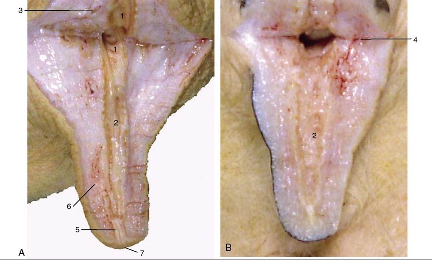
Figure 29-43 A, B, Section of a cow's teat and lactiferous sinus. 1, 2, Lactiferous sinus; 1, gland sinus; 2, teat sinus; 3, openings of lactiferous ducts; 4, submucosal venous ring; 5, papillary duct; 6, venous plexus in teat wall; 7, teat orifice.
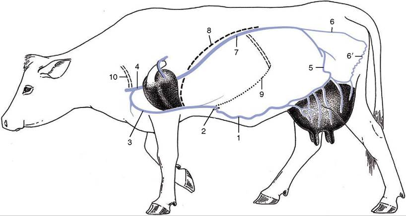
Figure 29-44 The venous drainage of the udder. 1, Subcutaneous abdominal (milk) v.; 2, milk "well"; 3, internal thoracic v.; 4, cranial vena cava; 5, external pudendal v.; 6, internal pudendal v.; 6', ventral labial v. (connecting ventral perineal v. with caudal mammary veins); 7, caudal vena cava; 8, diaphragm; 9, costal arch; 10, first rib.
At full term, the mammary glands exhibit short but well-formed teats, small sinuses, and the first branches of the duct systems. The bulk of the udder consists of fat. During the next few months growth keeps pace with the general growth of the body and is entirely due to deposition of fat. Thereafter, and thus commencing well before puberty, growth quickens; the rapid development of both the duct system and the gland tissue is probably due to the cyclical production of estrogen because spurts of activity occur directly before ovulation.
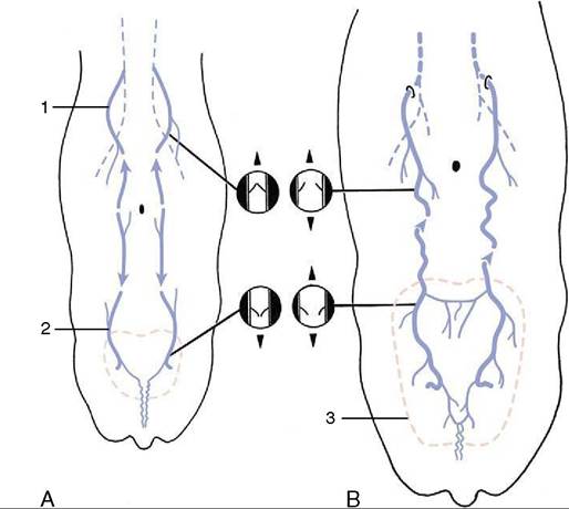
Figure 29-45 Development of the subcutaneous abdominal veins (schematic dorsal view). A, The region drained by the cranial superficial epigastric vein (1) is separated from that of the caudal superficial epigastric vein (2) in the calf and heifer. The valves in the cranial superficial epigastric vein direct blood cranially, while those in the caudal superficial epigastric vein direct blood caudally. B, The subcutaneous abdominal vein is formed during pregnancy. The increased blood flow through the enlarging udder (3) causes the veins to distend, the valves to become inefficient, and the two drainage regions to unite, which allows blood to flow in both directions.
Although a well-developed duct system is present by the time a heifer first conceives, additional growth of the ducts predominates in the first months of pregnancy; growth of the secretory tissue occurs in the second half.
Growth in late pregnancy is dependent on prolactin and growth hormone of hypophysial origin, in addition to progesterone and estrogen. Secretion of milk is later maintained by corticotropin, thyroid-stimulating hormone, and somatotropin. Regular milking is also necessary to maintain production. Since the act of milking stimulates the release of prolactin, oxytocin, and corticotropin, more frequent milking, within limits, increases the yield.
The mammary gland is composed of tubuloalveolar secretory units grouped to form lobules defined by connective tissue septa (Figure 29-48, A). The secretory alveoli are lined by a simple epithelium that changes markedly in height during the cycle of activity. The cells demonstrate maximal activity in those alveoli prepared to release milk when stimulated by suckling (or milking). After this the alveolar lumina are collapsed and irregu-
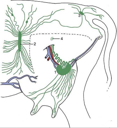
Figure 29-46 Lymph drainage of the udder. The broken line indicates where the left limb was removed to expose the udder. 1, Mammary (superficial inguinal) lymph node; 2, subil- iac lymph node; 3, ischial lymph node; 4, position of deep inguinal (iliofemoral) node.
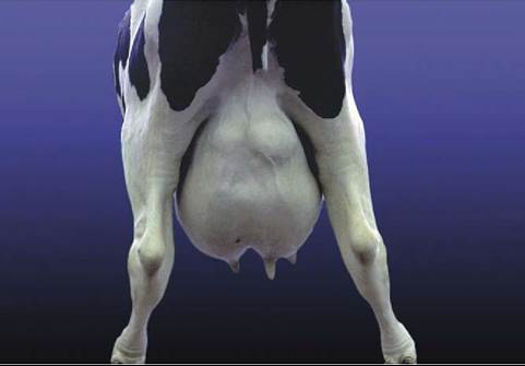
Figure 29-47 Holstein cow with enlarged mammary lymph nodes.
lar (Figure 29-48, B), and the epithelium is much reduced in height. All lobules within one gland do not necessarily exhibit the same stage of the secretory cycle, and both active and nonactive lobules may be present concurrently. The milk is forced from the secretory units into the duct system by contraction of surrounding myoepithelial cells (Figure 29-49). The interlobular and
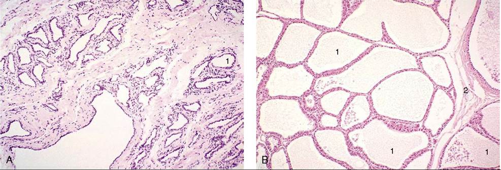
Figure 29-48 Section of (A) nonlactating and (B) lactating mammary glands; a compound tubuloalveolar gland (70?). 1, Alveolus; 2, interlobular septum.
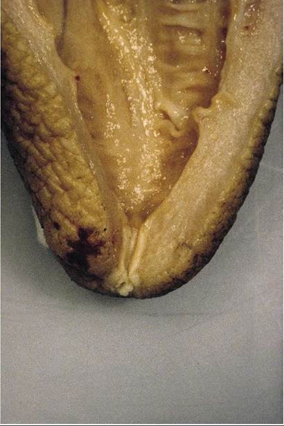
Figure 29-49 Section of the teat extremity showing the smooth muscle encircling the papillary duct.
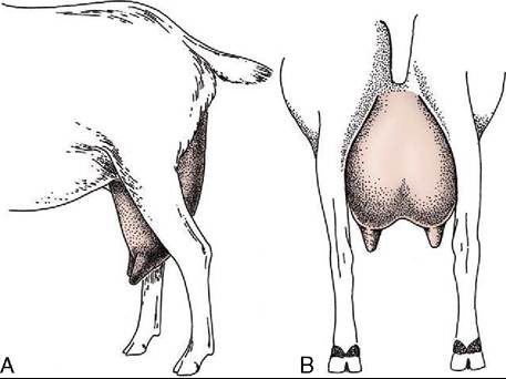
Figure 29-50 Lateral (A) and caudal (B) views of the goat's udder.
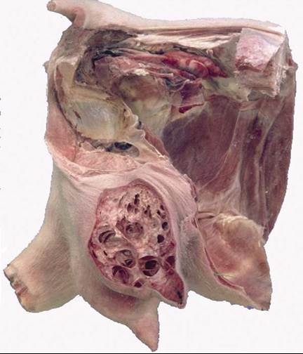
Figure 29-51 Sagittal section of a young goat's udder and teat.
intralobular connective tissue provides important structural support and conveys blood, lymph vessels, and nerves.
The udder of the small ruminants combines two glands that are more (goats) or less (sheep) distinctly demarcated externally. In milk goats the udder is large in relation to body size, deep, and conical (Figure 29-50); in ewes it is smaller and more hemispherical, although inclining toward the caprine form in breeds used for cheese production. The teats are cylindrical in the young, but in older animals, especially in goats of high productivity, they tend to become conical and blend more smoothly with the contours of the gland (Figure 29-51). Accessory teats are not uncommon in goats. The udder skin is finely haired in goats; in sheep the upper part may cover the fleece.
The structure, suspension, and vascular arrangements generally resemble those of the bovine udder. However the teats are not wholly naked. In sheep, closure of the papillary duct is achieved without the presence of a sphincter muscle.