» The Udder
The mammary glands are consolidated in a rather small udder situated below the caudal part of the abdominal floor and cranial part of the pelvis and concealed from casual inspection by the thigh (Fig.
22.25). The form and size of the udder vary with the present state and previous history of the mare; the udder is very small in young virgin animals. A prominent external groove indicates its formation from right and left halves; each half has the form of a laterally compressed cone and, though carrying a single teat, is composed of two (occasionally three) separate duct systems.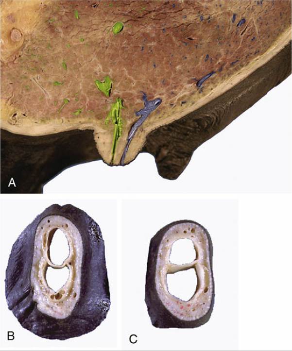
FIG. 22.26 (A) Sagittal section of the udder demonstrating the construction of the teat and the location of the lactiferous sinus. (B) and (C) Transected teats showing internal division.
The skin over the udder is thin, strongly pigmented, and sparsely haired. It glistens from the secretion of many sweat and sebaceous glands. The teat is small and cylindrical, except in the lactating mare, in which it is both larger and more conical. Two (or three) openings perforate the apex; each leads through a short papillary duct to a small lactiferous sinus spread between the teat and gland mass and associated with an independent set of lactiferous ducts (Fig. 22.26A-C). The tissues of the individual glands of each side interdigitate, and it is impossible to demonstrate their independence on dissection. Although much less developed, the suspensory apparatus resembles that of the cow's udder and combines medial elastic and lateral fibrous ligaments, which together encapsulate the udder and supply the lamellae that support the parenchyma. The medial ligaments provide a cleavage plane between the apposed surfaces of the udder halves.
The blood supply comes from the external pudendal artery, and the principal venous return is by the corresponding vein, which does not follow the usual course through the inguinal canal (p.
540). As in the cow, a subcutaneous venous connection with a superficial vein of the thoracic wall develops as an alternative drainage route during the first pregnancy. Lymph drains to the mammary (superficial inguinal) nodes. The cutaneous innervation is divided between the nerves of the flank and a descending (mammary) branch of the pudendal nerve; the contributing spinal nerves are thus those of cord segments L2-L4 and S2-S4 (Fig. 22.27). The substance of the gland is supplied by the genitofemoral nerve (L3-L4). The glands develop rapidly during the second half of the first pregnancy and commence secretion before birth. Sebaceous secretion, epithelial debris, and possibly colostrum that escape through the teat openings during the last days of pregnancy dry to give the apex a waxy covering, which is a useful indication that parturition impends.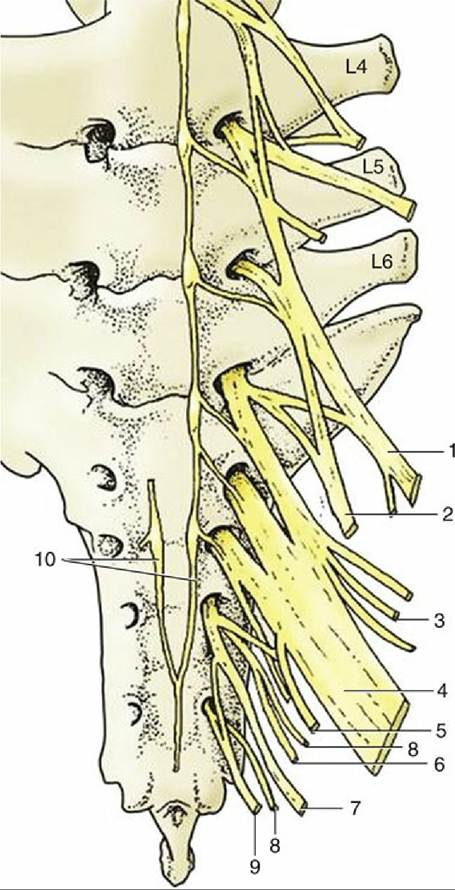
FIG. 22.27 Ventral view of sacrum and caudal lumbar vertebrae (L4-L6) with emerging ventral rami forming the lumbosacral plexus. 1, Femoral nerve (n.); 2, obturator n.; 3, cranial gluteal n.; 4, sciatic n.; 5, caudal cutaneous femoral n.; 6, caudal gluteal n.; 7, pudendal n.; 8, pelvic n.; 9, caudal rectal n.; 10, continuation of sympathetic cord.
Comprehension Check
As a group, compare the anatomy of the equine and ruminant female and male reproductive tracts, including the structure of the pelvis.
I =<
* These are terminal arteries that open directly into the cavernosal spaces of the erectile tissue of the penis. Their myoepithelial walls cause them to be coiled (helicine) and closed in the flaccid penis. Sexual stimulation relaxes them, which allows blood to engorge the erectile tissue.
The Forelimb of the Horse
In the Western world horses are now mainly bred for use in sport and recreation, pursuits that often make heavy demands on their speed and endurance and expose their limbs to continual strain and repeated risk of injury. Even relatively minor incapacity may unfit a horse for this work, and the importance of soundness of limb is crisply stated by the old adage "no foot, no horse." Considering the importance of lameness in equine medicine, a detailed knowledge of the anatomy of the limb is needed.
The limbs of the horse display extreme adaptations for fast running with a concomitant loss of versatility. The forelimbs carry the greater part (some 55%-60%) of the body weight at rest and supply the principal shock absorbers that are necessary in the faster gaits and especially when landing from a jump. The hindlimbs furnish the main propulsive thrust. Furthermore, the share of the load that is supported by each limb may be altered by varying the posture to shift the center of gravity. The most obvious maneuver is to raise or lower the head to displace the center caudally or cranially, respectively. The lame animal also lifts the head when a painful forelimb is placed on the ground and lowers it when the sound limb bears weight. Because the later movement is more obvious, a horse with forelimb lameness is said to "nod on the sound foot." When there is a painful condition of a hindlimb, the head is lowered as the affected limb assumes support.
A forelimb with good conformation is straight when viewed from the front. A line dropped from the point of the shoulder bisects the limb and passes through the center of the hoof; the digit continues the cannon (metacarpus) in a straight line, neither "toeing-in" nor "toeing-out" (Fig. 23.1). Much of the limb should also be straight when viewed from the side. A line dropped from the tuberosity of the scapular spine should bisect it to the fetlock and then pass just behind the hoof, whose slope should parallel that of the digit. Deviations from the normal conformation can result in abnormal movements, which in turn may cause interference between the feet, unequal and abnormal hoof wear, and development of lameness.
The more common deviations seen when viewing from the front are categorized as "base-wide," in which the limbs slope laterally, and "base-narrow," in which they slope medially. Deviations seen from the side include "standing under," in which the limbs slope caudally, and "camped," in which they slope cranially.
Cranial, caudal, medial, and lateral deviations of the carpus are also recognized; the last two faults are "knock-knees" and "bowlegs."Retention of the full length of the shaft of the ulna is a congenital anomaly that is fairly common in Shetland ponies. It is associated with a valgus deformity*-sometimes very severe—of the limb.
The distinctive "leggy" appearance of the young foal must be familiar to every reader (Fig. 23.2). The acquisition of the adult shape involves changes in the ratios of the lengths of the limbs (taken as a whole) to that of the trunk and in the ratios between the lengths of successive segments of the limbs—arm (thigh), forearm (leg), and metacarpus (metatarsus). According to one source, in the newborn Thoroughbred the ratio of the humerus (femur) to the metacarpus (metatarsus) is approximately 4:5 (4:5); in the adult the ratio is approximately 6:5 (6.5:5). These changes are achieved through a postnatal growth in length of the metacarpal (metatarsal) bones of about 20% and growth of the humerus and femur of about 100%.
The cutaneous features known as chestnuts and ergots are described on page 362 (see Fig. 10.17).