The Uterine Cornu
The uterus comprises the two uterine cornu (or horns), the uterine body and the cervix. The uterus receives the fertilised ovum or ova, which become blastocyst(s) before implanting in the endometrium of the uterine wall.
The developing embryo attaches to the uterus by a placenta (see Section 14.5), which allows the nutrients to pass from the maternal blood vessels to the blood of the foetus. The uterus comprises three layers. The outer layer is a serosa that is continuous with the visceral peritoneum and the mesometrium of the broad ligament. The middle layer (myometrium) consists of circular and longitudinal muscle fibres. The inner layer is the thick mucous membrane of the endometrium and includes a columnar epithelium and tubular glands.The uterus is suspended by the broad ligament (Section 3.4.3), a peritoneal fold that is attached to the dorsal abdomen and the walls of the pelvis. There are three divisions of the broad ligament, according to their uterine attachments, i.e. mesovarium, mesosalpinx and mesometrium. There is usually a substantial deposit of fat in the broad ligament.
Mammalian uteri are classified according to their structure. There are four main types of uterus as follows:
Simplex: The entire uterus is fused into a single organ. Found in primates including human beings.
Duplex: There are two separate uterine bodies and cervixes but a shared vagina and vestibule. Found in marsupials, rodents and rabbits.
Bipartite: There are two separate uteri for most of their length but there is a shared uterine body, cervix and vagina. Found in ruminants, horses and cats.
Bicornuate: The upper parts of the uterus remain separate but the rest of the uterus is fused as a single organ. Found in pigs, dogs, elephants and whales.
14.1.1 Species variations
Mare (Figure 14.1): The uterine cornua are arranged in a flattened T shape, the ovarian extremities of each cornu being rounded and blunt rather than tapered.
The cornua are relatively short compared with the other domestic species.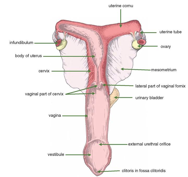
Figure 14.1 Dorsal view of the female reproductive organs of the mare. The ovaries have been rolled over so that the opening of the ovarian bursa is visible.
Since the placenta is diffuse (see Section 14.5), the mucous membrane is the same all over and contains many branched and coiled mucus-secreting glands. A localised region about the size and shape of a horseshoe and containing raised patches becomes apparent at about 25 days of gestation. These are the endometrial cups; they are about 3 cm in diameter with a central depression and disappear around halfway through pregnancy. The cells of the endometrial cups secrete equine chorionic gonadotrophin (equine luteinising hormone).
Isolated, acellular, oval objects may be present floating within the amnion or allantois. These are the hippomanes and occur in the ox, sheep and pig but most often in the horse. They are flattened and smooth. They are 2.5-10 cm in length and light brown in colour. Their significance is unknown, but it is suggested that they have aphrodisiac properties.
Cow (Figure 14.2): The uterine cornua are curled caudally so that the ovaries are close to the body of the uterus and the shaft of the ilium. The cornua gradually taper into the uterine tube. The length of the uterus is relatively long (35 cm).
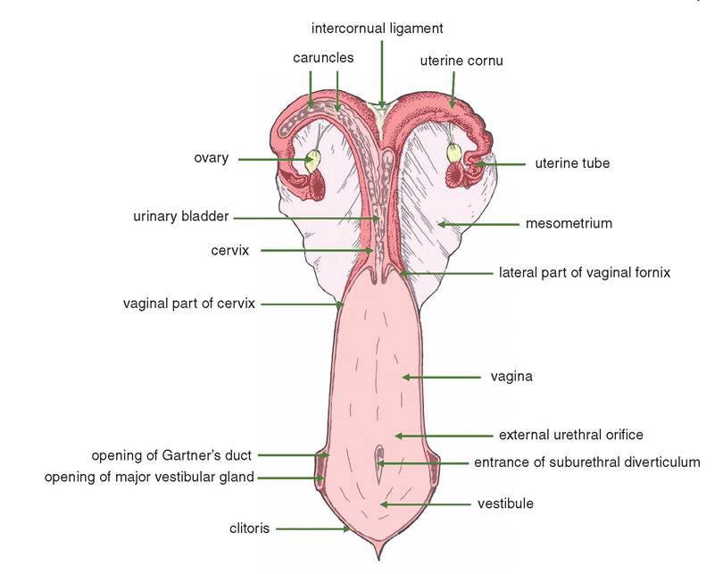
Figure 14.2 Dorsal view the reproductive organs of the cow. The ovaries have been rolled over so that the opening of the ovarian bursa is visible.
The placenta of the cow is cotyledonary (see Section 14.5) and consists of about 100 caruncles on the endometrium that form attachments with the foetal cotyledons. A single unit of a caruncle and a cotyledon is called a placentome. In the non-pregnant state the caruncles are inconspicuous oval knobs about 1.0 cm in diameter.
During pregnancy they can reach 10.0 cm in length and are attached to the uterine wall by a stalk containing their blood vessels. The removal of retained foetal membranes involves the risk of pulling off caruncles with inevitable haemorrhage. In the non-pregnant uterus the surface of the caruncles is smooth. In pregnancy the surface of the endometrium is pitted with a sponge-like network of deep crypts that accommodate the villi of the allantochorion (see Section 14.5).The intercornual ligament (Figure 14.2) is well developed in the ox and is usually double at its cranial end; it binds the diverging cornua together.
Ewe: The uterine cornua of the sheep resemble those of the ox, but they are smaller. Black pigmentation of the caruncles and uterine tubes is common but not always present; a small uterus of a calf with no pigmentation resembles that of a ewe. The pregnant
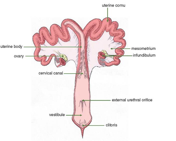
Figure 14.3 Dorsal view the reproductive organs of the sow. The ovaries have been rolled over so that the opening of the ovarian bursa is visible.
caruncles are circular with a deep depression in the centre. The intercornual ligament is single in the ewe.
Sow (Figure 14.3): The uterine cornua are very long in this species (or voidance to the maternal blood by the placenta.
14.3