THE UTERUS
The uterus has a large body and two divergent horns. The horns, which are about 25 cm long, lie wholly within the abdomen and diverge sharply from each other. They are suspended from the abdominal roof by the broad ligaments, whose width varies such that the extremities of each horn are more tightly tethered than the intermediate part (Figure 22-8/14).
However, in life, the horns are usually raised toward the abdominal roof on the mass of intestines. The body of the uterus is a little shorter (≈20 cm) than the horns and lies partly within the abdomen and partly within the pelvis. Although its relations vary, they always include the terminal part of the descending colon and rectum dorsally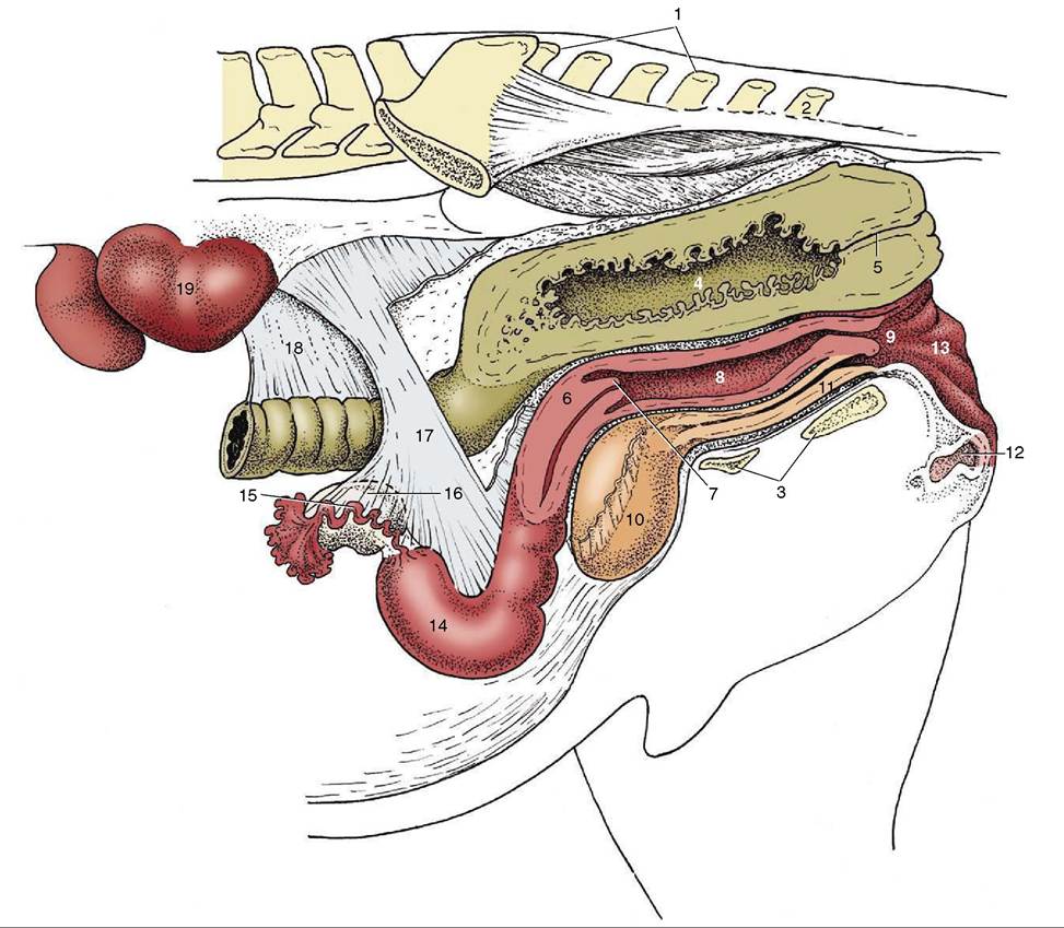
Figure 22-8 Caudal abdominal and pelvic organs of the mare in situ; the organs have been sectioned in a paramedian plane with the pelvis. Because of the absence of the intestines, the ovaries hang much lower than they would in the intact animal. 1, Sacrum; 2, Cd2; 3, floor of pelvis; 4, rectum; 5, anal canal; 6, cervix; 7, vaginal part of cervix; 8, vagina; 9, vestibule; 10, bladder; 11, urethra; 12, clitoris; 13, vulva; 14, left uterine horn; 15, uterine tube; 16, ovary; 17, broad ligament (largely cut away); 18, descending mesocolon; 19, left kidney.
and the bladder and various parts of the gut ventrally. The body is often displaced to one side by a distended bladder or by pressure from the gut. When the uterus is empty, both horns and body are flattened and the lumen almost obliterated.
The cervix (Figure 22-8/d) is rather short (≈6 cm). Although its position and extent are not readily distinguishable on visual inspection, they are at once revealed on palpation as the cervix has a somewhat firmer consistency. The difference is less pronounced at estrus. The caudal part of the cervix projects into the lumen of the vagina, where it is surrounded by an annular space (fornix) of more or less uniform depth. This intravagi- nal part (Figure 22-8/7) has a lobed appearance created by the extension through the external ostium of the mucosal folds lining the cervical canal. These folds continue onto the vaginal wall, where they gradually subside. Except at estrus and parturition the cervical canal is closed; however, it will still admit a finger on gentle probing (Figure 22-12).
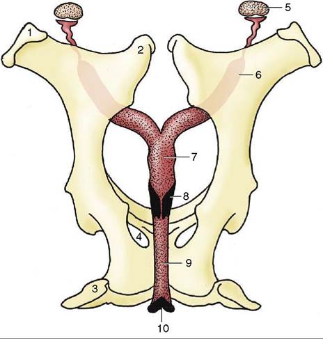
Figure 22-9 The female reproductive organs in relation to the pelvis, dorsal view. 1, Coxal tuber; 2, sacral tuber; 3, ischial tuber; 4, obturator foramen; 5, ovary; 6, uterine horn; 7, body of uterus; 8, cervix; 9, vagina; 10, vulva.