The Uterus
The uterus has a large body and two divergent horns. The horns, which are about 25 cm long, lie wholly within the abdomen and diverge sharply from each other. They are suspended from the abdominal roof by the broad ligaments, whose width varies such that the extremities of each horn are more tightly tethered than the intermediate part (Fig.
22.8/14). However, in life, the horns are usually raised toward the abdominal roof on the mass of intestines. The body of the uterus is a little shorter (≈20 cm) than the horns and lies partly within the abdomen and partly within the pelvis. Although its relations vary, they always include the terminal part of the descending colon and rectum dorsally and the bladder and various parts of the gut ventrally. The body is often displaced to one side by a distended bladder or by pressure from the gut. When the uterus is empty, both horns and body are flattened and the lumen is almost obliterated.The cervix (Fig. 22.8/6) is rather short (≈6 cm). Although its position and extent are not readily distinguishable on visual inspection, they are at once revealed on palpation because the cervix has a somewhat firmer consistency. The difference is less pronounced at estrus. The caudal part of the cervix projects into the lumen of the vagina, where it is surrounded by an annular space (fornix) of more or less uniform depth. This intravaginal part (Fig. 22.8/7) has a lobed appearance created by the extension through the external ostium of the mucosal folds lining the cervical canal. These folds continue onto the vaginal wall, where they gradually subside. Except at estrus and parturition the cervical canal is closed; however, it will still admit a finger on gentle probing (Fig. 22.12).
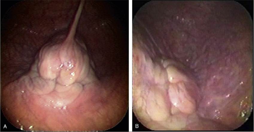
FIG. 22.12 The changing appearance of the cervix. (A) Diestrus. (B) Estrus.
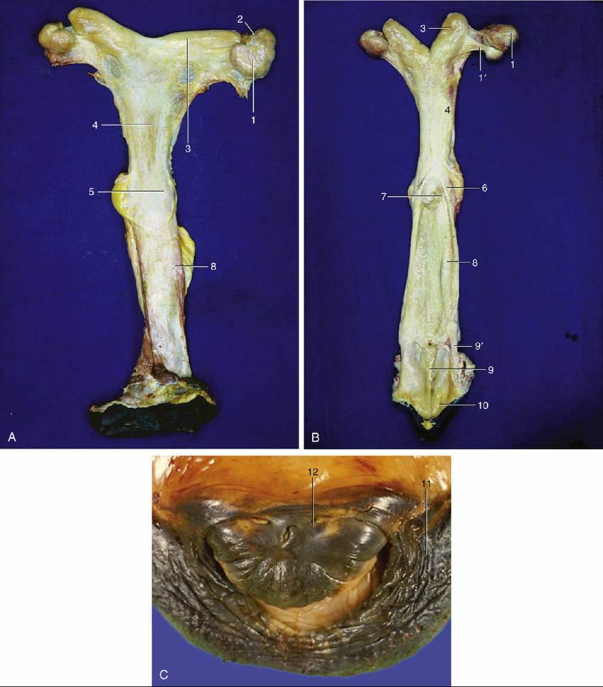
FIG. 22.13 (A) and (B) Dorsal view of the female reproductive organs. The dorsal wall of the caudal part of the tract has been opened in B. 1, Right ovary; 1', proper ligament of ovary; 2, uterine tube; 3, horn of uterus; 4, body of uterus; 5, cervix; 6, vaginal part of cervix; 7, fornix; 8, vagina; 9, vestibule; 9', wall of vestibule; 10, vulva. (C) An enlargement of the vulva, showing the glans of the clitoris within the ventral commissure. 11, Right labium; 12, glans of clitoris.