The Uterus
The sow's uterus is distinguished by its short body and long, tortuous horns (Fig. 35.4/5 and 8). The body, about 5 cm long, is deceptively short because the parts of the horns at their origin lie within common investments (as in ruminants).
In the nongravid state each horn measures about 1 m, and a fairly generous broad ligament (Fig. 35.4/6) gives it freedom of position, relations, and arrangement; however, it fails to reach the abdominal floor. Some parts become mingled with coils of small intestine and can be confused with these. The cervix, which lies half within the abdomen and half within the pelvis, is peculiar for its length (≈25 cm). The cervix is about 20 cm in length and has rows of interdigitating mucosal prominences (Fig. 35.4/11) that project into the lumen and close the canal, except at estrus and parturition. The cervix has many goblet cells that produce mucus during the estrus. Its junctions with the uterine body and the vagina taper and are ill defined.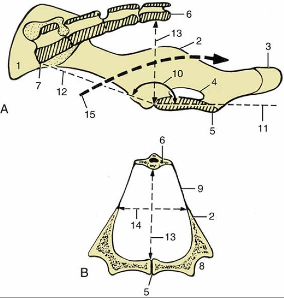
FIG. 35.1 (A) Median section of the sow's pelvis. (B) Transverse section of the pelvis near the level of the vertical diameter. 1, Coxal tuber; 2, ischial spine; 3, ischial tuber; 4, obturator foramen; 5, pelvic symphysis; 6, fourth sacral vertebra (S4); 7, promontory; 8, acetabulum; 9, sacrosciatic ligament; 10, angle between pelvic floor and conjugata; 11, plane of pelvic floor; 12, conjugata; 13, vertical diameter; 14, transverse diameter; 15, pelvic axis.
More on the topic The Uterus:
-
Veterinarian -