The Vagina
The remaining part of the genital tract is divided between the vagina and vestibule, approximately in the ratio of 3:1; the boundary is a few centimeters cranial to the ischial arch (Fig.
29.11). Because the vagina is capable of great expansion, in length and in diameter, its passive dimensions are not of great significance. The lining exhibits low folds, both circular and longitudinal, and the lumen is closed by the falling together of the roof and floor (Fig. 29.8). It is usual to find the caudal part ventrally narrowed, especially in young animals, because of the urethralis muscle.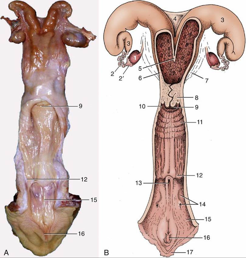
FIG. 29.14 The bovine reproductive organs, dorsal view. (A) The vagina and the vestibule have been opened in the specimen. (B) The greater part of the tract is shown opened in the schema. 1, Ovary; 2, uterine tube; 2', infundibulum; 3, uterine horn; 4, intercornual ligament; 5, wall of uterus dividing the two horns; 6, body of uterus with caruncles; 7, broad ligament; 8, cervix; 9, vaginal part of cervix; 10, fornix; 11, vagina; 12, position of former hymen; 13, external urethral orifice and suburethral diverticulum; 14, major vestibular gland and its excretory orifice; 15, vestibule; 16, glans of the clitoris; 17, right labium.
The cranial two-thirds of the dorsal wall faces into the rectogenital pouch, but caudal to this the vagina and rectum are joined by a wedge of tissue (Fig. 29.11). The ventral surface has a less complete peritoneal covering and is related to the bladder and urethra and to the packing tissues about the urethra. The lateral walls are also largely without peritoneum, being cranially included in the broad ligament and more caudally sharing in the general retroperitoneal arrangement (Figs. 29.7 and 29.8). This limitation of the peritoneum is relevant to the prognosis of wounds to the vaginal wall. The peritoneal covering of the dorsal fornix region provides a convenient route for surgical access to the abdominal cavity, most often used for operations on the ovary; it has the additional advantage of avoiding the major vessels that pass below and to the sides of the vagina.
Vestiges of the mesonephric ducts may be found below the mucosa of the floor near the junction with the vestibule; they are sometimes the origin of cysts.
The vagina is almost absent in the freemartin (p. 701), whose abnormally short tract is evident on examination of the vestibule. Aplasia or constriction of the vagina also occurs in white heifer disease, another congenital anomaly. The freemartin is found after a twin pregnancy in which the female fetus is adversely affected by the male twin (Fig. 29.18).