The Vagus Nerve (X)
The vagus nerve is the nerve of the fourth and subsequent pharyngeal arches. It also contains the parasympathetic fibers that innervate the cervical, thoracic, and abdominal viscera.
The second component gives it by far the most widespread distribution of any cranial nerve (Fig. 8.72/5).The vagus forms part of the bundle of nerves that passes through the jugular foramen. It bears two small ganglia on the stretch that lies within and immediately external to the foramen, and beyond this the vagus runs in close association with the glossopharyngeal and accessory nerves. After the glossopharyngeal nerve turns rostrally, the vagus continues caudally, running close to the cranial cervical ganglion. It then continues down the neck in close contact with the sympathetic trunk, with which it is bound within a common fascial sheath, on the dorsal margin of the common carotid artery, alongside the trachea. The left vagosympathetic trunk has an additional contact with the esophagus. The vagus and sympathetic nerves diverge at the entrance to the chest, after which the vagus continues more or less horizontally through the mediastinum until it divides over the pericardium into dorsal and ventral branches. These branches combine with the corresponding contralateral branches to form the dorsal and ventral vagal trunks that enter the abdomen along the corresponding borders of the esophagus. Within the abdomen, the two nerves branch freely, participating with the sympathetic fibers in forming the plexuses from which the abdominal viscera are supplied (p. 315).
The first significant branch from the main trunk after the vagus nerve leaves the skull is an auricular branch that takes part in the innervation of the skin of the external ear. This is followed by pharyngeal branches that combine with those of the glossopharyngeal, cranial laryngeal, and sympathetic nerves in forming the pharyngeal plexus.
An extension of the plexus supplies the cervical esophagus. The cranial laryngeal nerve goes to the larynx, where it divides into an external branch for the cricothyroid muscle and an internal branch for the laryngeal mucosa from the aditus to the glottis. This internal branch makes connections with the recurrent laryngeal nerve (described later). The depressor nerve to the heart is formed partly of fibers from the cranial laryngeal nerve and partly of fibers from the main vagal nerve; it is difficult to follow because in most animals it rejoins the main trunk for its further progress through the neck and thorax to the heart.The thoracic portion of the vagus detaches cardiac branches that form a mediastinal plexus with sympathetic fibers also innervating cardiac muscle. A large caudal (recurrent) laryngeal nerve is also detached within the thorax. The recurrent laryngeal nerve of the right side changes direction by winding around a branch of the subclavian artery, and the left one winds around the aorta. The recurrent laryngeal nerve reascends the neck ventral to the common carotid artery in a course that leads it back to the larynx, where it supplies the bulk of the intrinsic laryngeal musculature (all but the cricothyroideus) and the mucosa caudal to the glottis. Small twigs from the recurrent laryngeal nerve detached en route pass to the cardiac plexus and to the trachea and esophagus. The distribution of the main trunk is completed by pulmonary branches that combine in a common plexus with sympathetic nerves.
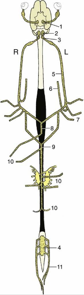
FIG. 8.72 Origin and distribution of the parasympathetic nervous system. Ventral view, schematic. 1, Parasympathetic oculomotor nucleus; 2, rostral and middle parasympathetic nuclei of the medulla oblongata; 3, dorsal vagal nucleus; 4, sacral outflow; 5, vagus nerve; 6, recurrent laryngeal nerve; 7, parasympathetic fibers to heart and lungs; 8, ventral vagal trunk; 9, dorsal vagal trunk; 10, parasympathetic fibers to the abdominal organs; 11, pelvic nerves; L, left; R, right.
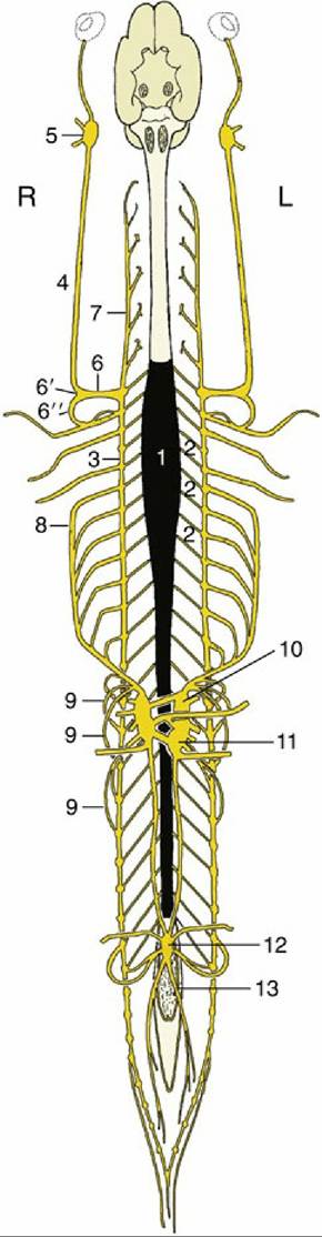
FIG. 8.73 Origin and distribution of the sympathetic nervous system. Ventral view, schematic. The parasympathetic nuclei in brain and spinal cord are indicated in gray. 1, Sympathetic outflow from T1 to L3; 2, communicating branches; 3, and 4, sympathetic trunk; 5, cranial cervical ganglion; 6, cervicothoracic ganglion; 60, middle cervical ganglion; 62, ansa subclavia; 7, vertebral n.; 8, greater splanchnic n.; 9, lesser splanchnic nn.; 10, celiac ganglion; 11, cranial mesenteric ganglion; 12, caudal mesenteric ganglion; 13, hypogastric n.
Damage to the vagus nerve and its branches may be manifested in a variety of ways, including difficulties in swallowing and altered functioning of the heart and the other viscera. Degeneration of the recurrent laryngeal nerve is especially common in horses, producing the condition known as roaring (p. 515); it also occurs in dogs.