THE VENTRAL PART OF THE NECK
Dorsal cervical structures are described with the vertebral column (Chapter 26). The skin of the ventral aspect is freely movable and redundant in amount; it becomes folded and creased when the head is lowered to the ground.
In addition, the caudal part of the neck carries the large dewlap that continues onto the brisket (breast) between the forelimbs (Figure 25-22). There is scant evidence for the belief that this increase in surface area is important in heat dissipation as is sometimes claimed, for the Zebu in particular. Zebu cattle do possess, here and elsewhere, more numerous, larger, and more saclike sweat glands than are found in cattle of European origin.The groove over the course of the external jugular vein is generally obvious, at least in cows. It is bounded dorsally by the brachiocephalicus (cleidomastoideus) extending from the arm to the skull and ventrally by the part (sternomandibularis) of the sternocephalicus that runs between the manubrium of the sternum and the angle of the jaw. Except in the most caudal part of the neck, a second part of the sternocephalicus (sternomas- toideus) forms the floor of the groove and provides a substantial separation between the vein and the common carotid artery (Figure 25-23/7). The external jugular vein is easily raised for injection and blood sampling because only the caudal part is covered by the cutaneous muscle, and even this is rather weak. The vein is formed caudal to the parotid gland by the confluence of maxillary and linguofacial radicles (see Figure 25-2). It is the principal drainage of the head and neck but is assisted by the internal jugular vein, the vertebral vein, and the internal vertebral plexus. Variation in the prominence of the vein may reflect conditions within the thorax. Gentle undulation in time with respiration is due to a change in intrathoracic pressure.
Pulsation in time with the heartbeat in healthy cattle indicates the recurrence of atrial systole; in other animals it points to atrioventricular valvular incompetence. The normal jugular pulse does not persist after compression of the cranial part of the vein, but the pathological pulse does.The superficial muscles enclose the space that contains the cervical viscera and the vessels and nerves that make their way between the thorax and the head (see Figure 25-23). All of these organs are invested by tough fascia and are joined by looser tissue.
The trachea may be identified on deep palpation and is most easily appreciated toward the upper end of the neck, between the diverging sternocephalic muscles; even here it is not directly subcutaneous because the thin straplike sternothyrohyoid muscles follow its whole length. The trachea (Figure 25-23/14) is small in section and slightly deeper than it is wide; its form makes it
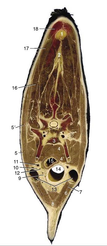
Figure 25-23 Transverse section through the middle of the bovine neck. 1, 2, Nuchal ligament (funiculus and lamina nuchae); 3, vertebra; 4, longus colli; 4', longus capitis; 5, 5', brachiocephalicus; 5, cleidooccipitalis; 5', cleidomastoideus; 6, 7, sternocephalicus; 6, sternomandibularis; 7, sternomastoi- deus; 8, combined sternohyoideus and sternothyroideus; 9, thymus and internal jugular vein; 10, recurrent laryngeal nerve; 11, common carotid artery; 12, vagosympathetic trunk; 13, external jugular vein; 14, trachea; 15, esophagus; 16, omotransversarius; 17, trapezius; 18, rhomboideus.
susceptible to narrowing by local pressure. The symmetry of its relations is disturbed by the devious course of the esophagus. Its structure is mainly remarkable for the concentration of lymphoid tissue in the dorsal ret- romucosal space (external to the tracheal muscle but within the cartilage rings).
Although the esophagus cannot be identified by palpation, its position is made evident by the swift movement along its track when the animal swallows.
In its cervical course the esophagus gradually slips to the left of the trachea only to creep back to a more dorsal position as the thorax is approached. However, its position varies with posture; its course is considerably straightened when the neck is extended. The relations in the middle of the neck are shown in Figure 25-23.The ruminant esophagus is very distensible, but its wide appearance in the cadaver gives a misleading impression of the usual condition in life. The mucosa is remarkably insensitive, which is one reason why cattle rarely appear to be distressed by the passage of a stomach tube or probang. Although transport is normally rapid in both directions, chunks of food quite commonly become lodged in the esophagus. The predilection sites are at the origin from the pharynx, at the thoracic inlet, and level with the tracheal bifurcation.
The thyroid gland is almost completely divided into two lobes, each shaped like an inverted pyramid and placed laterally over the cricoid cartilage. They are tenuously joined by an isthmus that crosses the second tracheal ring ventrally. They are finely granular and brick-red in the adult but paler in the calf (see Figure 6-4, C).
The parathyroid glands are small (ca. 8 to 10 mm) and, because they are irregular in shape and inconstant in position, frequently difficult to find. They may be embedded in other structures—usually the thyroid, thymus, or mandibular gland. The external parathyroid most often lies cranial to the thyroid but caudal to the carotid bifurcation; the internal one is perhaps most often embedded in the thyroid or located between this and the trachea. They have been confused with lymph nodes, which they resemble superficially.
The thymus is large and lobulated and extends from the larynx to the pericardium in young animals (Figure 25-24/1,2). Its cervical part is connected to the thoracic thymus by a narrow isthmus ventral to the trachea. The cervical part comprises two horns that taper over the lateral aspects of the trachea, possibly reaching the larynx; the cranial tip may be, or appear to be, detached and fragmented and more closely associated with the medial retropharyngeal lymph node and the mandibular and parathyroid glands.
The thymus grows rapidly during the first 6 or 9 months of postnatal life, although it attains its greatest relative size much earlier. Indeed, involution may begin as early as the 8th week after birth. The tempo of regression varies, and the thymus, particularly its thoracic part, may still be quite large in animals several years old. Ultimately the isthmus and neck part disappear almost completely. The thymus of young calves is bright pink or even red, but the organ lightens with age; its consistency also firms as the active tissue is progressively replaced by fatty fibrous tissue.The common carotid artery runs dorsolateral to the trachea within a fascial sheath shared with the vagosympathetic trunk. The internal jugular vein and the
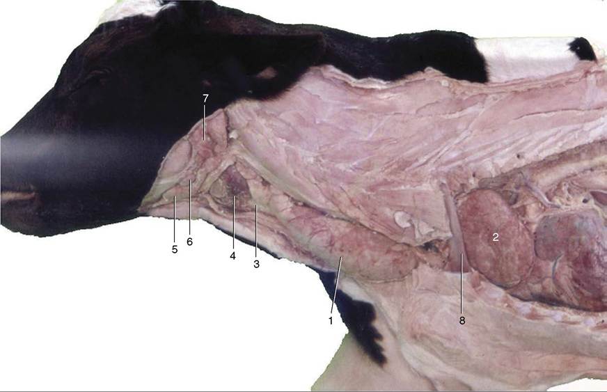
Figure 25-24 The thymus in the newborn calf. 1, Cervical part of thymus; 2, thoracic part of thymus; 3, trachea; 4, thyroid gland; 5, mandibular gland; 6, mandibular lymph node; 7, parotid gland; 8, first rib.
recurrent laryngeal nerve are closely related to the sheath on the right side; the esophagus intervenes on the left. The artery ends over the lateral pharyngeal wall, where it detaches a small occipital artery; the parent trunk is continued (without alteration of course) as the external carotid artery. In the fetus an internal carotid artery arises with the occipital artery, but the part proximal to the rete mirabile (see Figure 7-35) begins to close even before birth; complete obliteration is usually achieved a few months after birth, although a residual lumen sometimes persists for a year or two (Figure 25-25/4). The common carotid artery detaches no branches of individual consequence before its termination. Pulsation in the common carotid may sometimes be detected when the artery is pressed against the transverse processes of the vertebrae.
At this point brief mention may be made of the blood supply to the brain, less because of any clinical significance than on account of its relevance to the controversial Jewish and Muslim slaughter techniques, in which the animals are killed by a deep ritual slash of the neck without preliminary stunning.
The brain is supplied by a combination of vessels that feed very intricate arterial plexuses within the cranial cavity, external to the dura mater and submerged within the cavernous and associated venous sinuses. These plexuses, the retia mirabilia, are formed by many closely wound, anastomosing arteries. The retia are entered on their peripheral aspect from several sources (see Figure 7-35); on the distal or cerebral side the network narrows to one emissary trunk that pierces the dural membrane to form the cerebral arterial circle with its fellow. The circle lies on the ventral aspect of the brain and gives off branches according to the conventional pattern. The basilar artery, which runs caudally over the medulla and continues down the spinal cord, is a contributor to the circle in cattle but leads blood from it in sheep. Although difficult to explain on hemodynamic grounds, all parts of the bovine brain are supplied by a mixture of carotid and vertebral blood, whereas in sheep the vertebral blood is restricted to the caudal part of the brainstem. These differences are germane to the ritual slaughter technique because the vertebral arteries are spared when the common carotid trunks are severed. The suggestion that abrupt reduction of the pressure within the cerebral arteries produces almost immediate loss of consciousness has been questioned.
The vagosympathetic trunk exhibits no particular features of note. The vagus and sympathetic components loosen their association and part company before entering the thorax. Their further courses and connections are described elsewhere. The recurrent laryngeal nerves resemble those of other species.

Figure 25-25 Branching of the left common carotid artery. 1, Common carotid a.; 2, occipital a.; 3, ascending palatine a.; 4, remnant of internal carotid a.; 5, medial meningeal a.; 6, external carotid a.; 7, linguofacial trunk; 8, lingual a.; 9, facial a.; 10, deep lingual a.; 11, sublingual a.; 12, submental a.; 13, inferior labial aa.; 14, superior labial a.; 15, infraorbital foramen; 16, caudal auricular a.; 17, masseteric branch; 18, superficial temporal a.; 19, transverse facial a.; 20, cornual a.; 21, maxillary a.; 22, inferior alveolar a.; 23, mental a.; 24, rostral and caudal branches to rete mirabile; 25, malar a.; 26, angular a. of the eye; 27, caudal lateral nasal a.; 28, dorsal nasal a.; 29, infraorbital a.; 30, sphenopalatine a.; 31, major and minor palatine aa.