» The Ventral Part of the Neck
The ventral part of the neck contains the visceral space occupied by the esophagus, trachea, and other structures passing between the head and the thorax. This space is bounded dorsally by the muscles below the vertebrae and laterally and ventrally by flatter muscles united by stout fasciae.
The foremost lateroventral muscles are the brachiocephalicus and sternocephalicus, which bound the groove occupied by the (external) jugular vein (Fig. 18.38/12). The caudal part of this groove is covered by the cutaneous muscle of the neck, which radiates from a manubrial origin; the muscle thins as it passes from its origin, which increases the prominence of the cranial part of the vein, the obvious target when the vein is raised for puncture (Fig. 18.39/9 and 11). The brachiocephalicus is described on p. 574.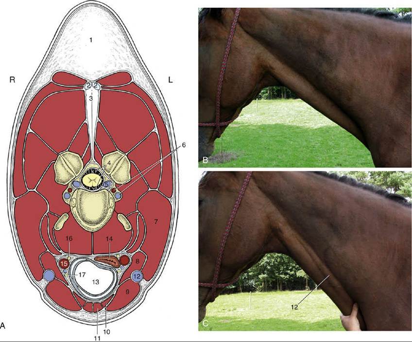
FIG. 18.38 (A) Transection of the neck at the level of the fourth cervical vertebra. L, Left side; R, right side. (B) and (C) The external jugular vein is not visible (B), but it is raised (C) when occluded in the jugular groove. 1, Crest; 2 and 3, funicular and laminar parts of nuchal ligament, respectively; 4, subarachnoid space; 5, internal vertebral venous plexus; 6, vertebral artery and vein; 7, brachiocephalicus; 8, omohyoideus; 9, sternocephalicus; 10, sternothyroideus; 11, sternohyoideus; 12, external jugular vein; 13, trachea; 14, esophagus; 15, common carotid artery; 16, vagosympathetic trunk; 17, recurrent laryngeal nerve.
The right and left sternocephalicus muscles arise from the manubrium side by side but diverge toward their mandibular insertions (Fig. 18.39/8). This leaves a median space through which the trachea may be palpated, although it is still covered by the thin sternothyroideus and sternohyoideus (Fig. 18.39/6). These are combined at their origin from the sternum but branch into slips that diverge to attach to the thyroid cartilage and the basihyoid.
The omohyoideus (Fig. 18.39/7), which extends between the medial aspect of the shoulder and the basihyoid, forms the floor of the jugular groove. It is said, unconvincingly, to protect the more deeply placed common carotid artery in unskillful venipuncture (Fig. 18.38/15). The muscles ventral to the trachea constitute the "strap muscles" that are resected in Forsell's operation for cribbing, which is a condition of stabled horses in which a horse hangs onto the crib with its teeth and dilates the pharynx to swallow air.FIG. 18.39
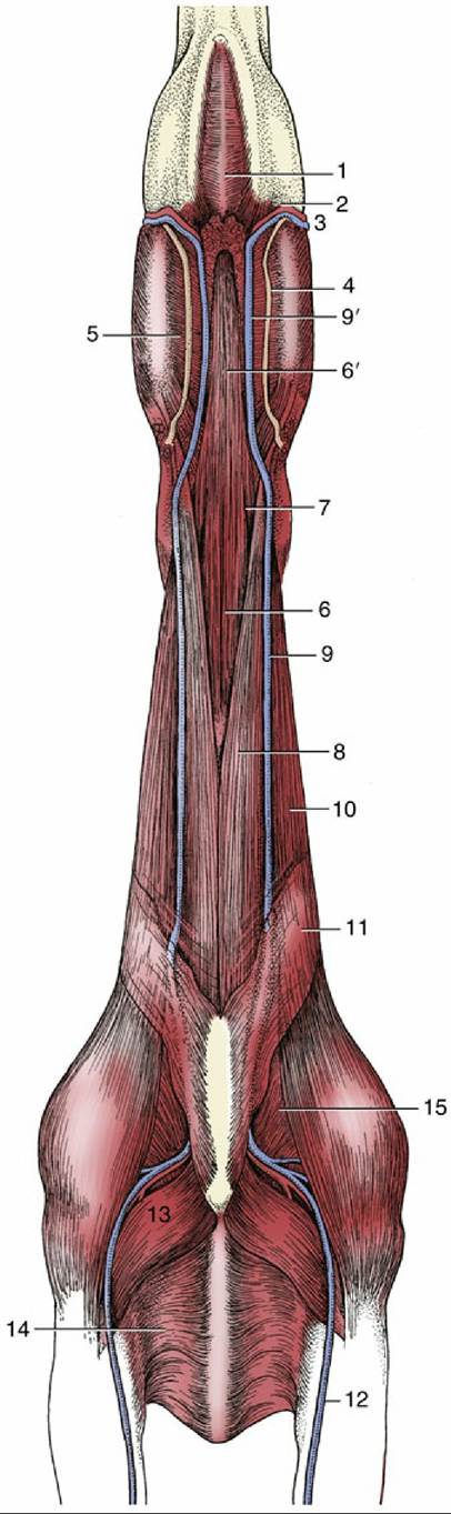
Ventral view of the neck and Intermandibular space. 1, Mylohyoideus; 2, mandibular lymph
nodes; 3, facial artery and vein; 4, parotid duct; 5, medial pterygoid; 6, sternohyoideus and sternothyroideus; 6', combined sternohyoideus and omohyoideus; 7, omohyoideus; 8, Sternocephalicus; 9, external jugular vein; 9', linguofacial vein; 10, brachiocephalicus; 11, cutaneous colli; 12, cephalic vein; 13, pectoralis descendens; 14, pectoralis transversus; 15, subclavius.
The trachea occupies a median position in the visceral space. Its size bears no constant relation to that of the body, which is an important point when selecting an endotracheal tube because the generous size of the glottis is not a limiting factor. The tracheal lumen is slightly flattened dorsoventrally and is of course maintained patent by the tracheal rings. It is therefore customary to completely transect as few cartilages as possible to avoid collapse of the wall in tracheotomy operations.
The esophagus begins dorsal to the trachea but slips to the left side by the middle of the neck (Fig. 18.38/14). It then slowly creeps back toward a median position, though it is often ventral to the trachea just before it enters the chest. It takes a more direct course when the neck is extended. The esophagus is too soft to identify easily on palpation, but its position is revealed when the animal swallows.
The common carotid artery lies ventral to the trachea at the base of the neck but gradually ascends to a more dorsal position (Fig. 18.38/15). It divides above the pharynx into the occipital, internal carotid, and external carotid arteries. The internal carotid supplies the brain, and the occipital supplies the region of the poll. The overall pattern of distribution of the external carotid artery is shown in Fig. 18.40. Pulsations of the common carotid may sometimes be felt in the middle of the neck when the artery is pressed against the subvertebral muscles. Nowadays, puncture at this site may be employed for the provision of a sample of arterial blood. The artery is enclosed in a thick fascial sheath shared with the vagosympathetic trunk, which follows its dorsal border. The recurrent laryngeal nerve lies ventral to it in the tracheal fascia (Fig. 18.38/16 and 17).
The deep cervical lymph nodes are scattered in packets—cranial, middle, and caudal—along the course of the tracheal lymph duct. The caudal group receives the outflow from the superficial cervical nodes (Fig. 18.41).
The external jugular vein is supplemented by the vertebral vein and the plexus within the vertebral canal in the drainage of the head. It is formed at the caudoventral angle of the parotid gland by the confluence of maxillary and linguofacial veins. It stands out very prominently and very conveniently for injection and sampling when raised by pressure over the jugular groove.
The lobes of the thyroid gland can be recognized on palpation as soft ovoid structures placed dorsolateral to the first part of the trachea (Fig. 18.26/21). They are joined ventrally by a narrow isthmus.
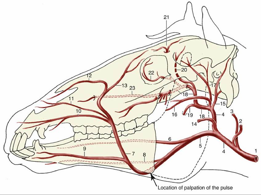
FIG. 18.40 Principal arteries of the head (schematic). 1, Common carotid artery (a.); 2, occipital a.; 3, internal carotid a.; 4, external carotid a.; 5, linguofacial a.; 6, lingual a.; 7, facial a.; 8, sublingual a.; 9, inferior labial a.; 10, superior labial a.; 11, lateral nasal a.; 12, dorsal nasal a.; 13, angularis oculi a.; 14, masseteric a.; 15, caudal auricular a.; 16, transverse facial a., displaced ventrally for clarity; 17, superficial temporal a.; 18, maxillary a.; 19, inferior alveolar a.; 20, caudal deep temporal a.; 21, supraorbital a.; 22, malar a.; 23, infraorbital a.
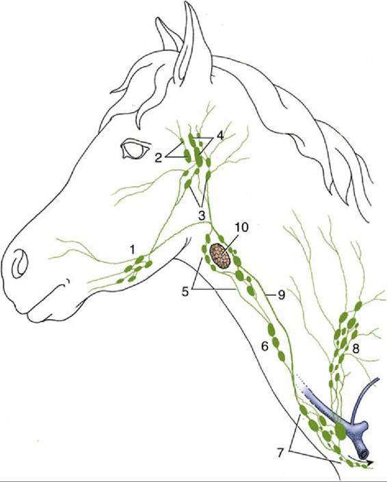
FIG.
18.41 Lymphatic structures of the head and neck (schematic). 1, Mandibular lymph nodes; 2, parotid lymph nodes; 3, medial retropharyngeal lymph nodes; 4, lateral retropharyngeal lymph nodes; 5, 6, and 7, cranial, middle, and caudal deep cervical lymph nodes, respectively; 8, superficial cervical lymph nodes; 9, tracheal duct; 10, thyroid gland.Although rarely as well developed as in the calf, a cervical part of the thymus may extend beside the trachea in the caudal part of the neck of the foal. It is often separated from the thoracic part and may be broken into several masses.