THE VERTEBRAL CANAL
ued behind the withers as the narrower supraspinous ligament. The second (laminar) part forms a fenestrated sheet closely applied to its fellow. It fills the space between the funicular part and the cervical vertebrae and consists of bundles of elastic fibers that run cranio- ventrally from the funicular part and the spines of T2 and T3 to attach to C2-C7.
Synovial bursae are interposed between the funicular part and certain bony saliences to minimize pressure. One, the nuchal bursa, is constantly present above the dorsal arch of the atlas; a second is sometimes found above the spine of the axis; and a third, the supraspinous bursa, is constantly present over the most prominent processes of the withers (Figure 19-3Z2,2',2"). Infections of the first and third, leading to conditions known as “poll evil” and “fistulous withers,” respectively, were formerly frequent and required extensive surgery for their eradication.The complicated arrangement of the powerful epaxial muscles of the back and neck conforms, but The relationships of the segments and cervical and lumbar swellings of the spinal cord to the vertebrae are shown in Figure 8-15. The first three sacral segments occur within the last lumbar vertebra, and the cord terminates within the cranial quarter of the sacrum of the adult (Figure 19-4).
The meninges remain separate to a more caudal level than in other species, and there is still a substantial subarachnoid space at the lumbosacral level. A communication exists in this species between the lumbar part of the space and a local widening (ventriculus terminalis) of the central canal of the spinal cord.
Both lumbosacral and caudal sites of injection are commonly employed to obtain epidural anesthesia. The procedure at the former level utilizes the divergence of the spinous process of the last lumbar and first sacral vertebrae for identification of the injection site (see Figure 19-4).
Although the interarcuate space is quite large, its distance (8 to 10 cm) from the skin makes it relatively easy to miss. “Low” epidural anesthesia is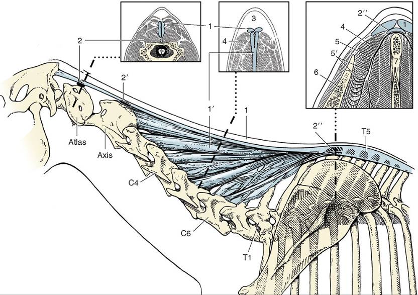
Figure 19-3 The nuchal ligament and associated bursae in lateral view and in three transverse sections. 1,1', Funicular and laminar parts of nuchal ligament; 2,2',2", cranial nuchal, caudal nuchal (inconstant), and supraspinous bursae; 3, fatty "crest" dorsal to nuchal ligament; 4, rhomboideus; 5, dorsoscapular ligament connecting spinous processes of the withers with the scapula; 5', elastic part of 5; 6, scapula; 7, spinous processes.
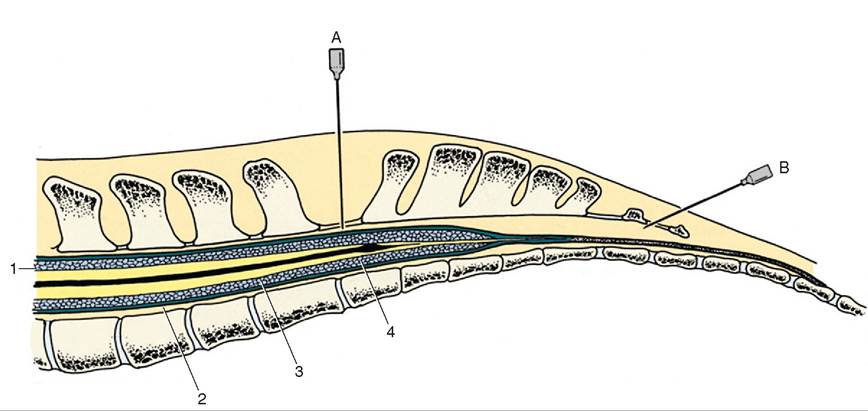
Figure 19-4 Median section of the equine vertebral canal and spinal cord. The lumbosacral interarcuate space and the space between the first and second caudal vertebrae are indicated by hypodermic needles placed for lumbosacral fluid collection (A) and for epidural anesthesia (B) for epidural anesthesia. 1, Pia mater; 2, dura mater; 3, arachnoid; 4, ventriculis terminalis.
performed between the first and second caudal vertebrae; the joint between these bones is very mobile, and the site for injection is readily discovered by “pumping” the tail up and down. The needle is inserted with a cranial inclination so that its point enters the canal within the first tail vertebra.
The vascularization of the spinal cord appears to be relevant to the etiology of a relatively frequent form of ataxia (“wobbles”) that occurs in foals and young horses. This may have its origin in congenital maldevel- opment and subsequent exostoses of the cervical articular processes that narrow the vertebral canal at the intervertebral levels. These exert pressure on the cord, although it is said that the cord lesions are secondary to interference with the venous drainage. In this context it should be known that the spinal arteries and veins are arranged in two sets, connected by relatively ineffectual anastomoses. One set enters the cord by way of the ventral fissure and supplies (and drains) the central gray substance and a thin surrounding shell of white. The second clambers over the lateral aspect to detach branches at intervals; these enter to supply (and drain) the bulk of the white matter (Figure 19-5). It is the veins of the second set that are supposedly compressed, leading to venous congestion and subsequent degeneration of the nervous tissue. It is claimed that the condition may develop in the fetus.
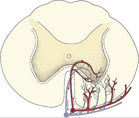
Figure 19-5 Blood circulation in the ventral part of the spinal cord, schematic. The blood supply to the gray substance and to the adjacent layer of the white is more or less independent of that to most of the white substance.