THE VERTEBRAL COLUMN
The vertebral axis runs parallel to the skin line in the loins and caudal part of the back; however, more crani- ally it is deflected ventrally. It reaches its lowest level at the entrance to the thorax; an abrupt flexure there places it on a path that gradually returns closer to the dorsal border as it ascends the neck (see Figure 26-1).
The vertebral skeleton and articulations follow the usual pattern, and few features need be mentioned. The vertebral formula is C7, T13, L6, S5, Cd18-20 in cattle; C7, T13, L6(7), S4 in sheep or S5 in goats; and Cd16-18 in both small ruminants. The great mobility of the neck allows the animal to raise and lower its head and to reach its side with its tongue. Most cervical movements represent the summation of small changes at several joints, but the adoption of the grazing position requires a considerable straightening at the cervicothoracic joint, where the neck vertebrae are brought into line with those of the chest. Although movements of the thoracic region are limited by the presence of the rib cage, the greatest flexibility of the trunk is found cranial to the level of the diaphragm. Behind this, movement is greatly
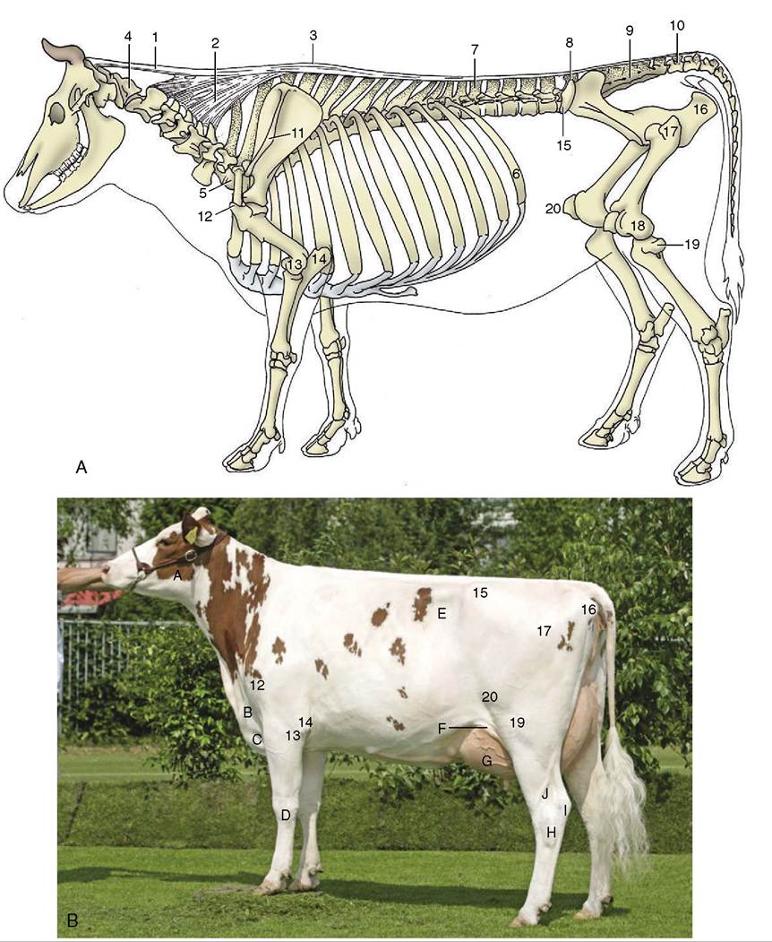
Figure 26-1 A, Skeleton with nuchal and supraspinous ligaments; most labeled parts are palpable. B, Cow in good condition. 1,2, Nuchal ligament; 1, funiculus nuchae; 2, lamina nuchae; 3, supraspinous ligament; 4, atlas; 5, last cervical vertebra (C7); 6, thirteenth rib; 7, first lumbar vertebra (L1); 8, last lumbar vertebra (L6); 9, sacrum; 10, first caudal vertebra; 11, spine of scapula; 12, greater tubercle; 13, 14, palpable features at elbow joint; 13, lateral epicondyle; 14, olecranon; 15, coxal tuber; 16, ischial tuber; 17, greater trochanter; 18, 19, 20, palpable features of stifle joint; 18, lateral condyle of femur ; 19, lateral condyle of tibia and remnant of fibula; 20, patella.
A, Masseter; B, jugular vein; C, brisket; D, carpus; E, paralumbar fossa; F, flank fold; G, udder; H, hock joint; I, calcaneus (point of the hock); J, lateral saphenous vein.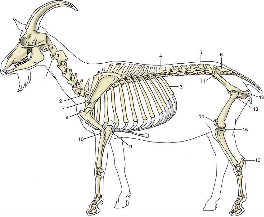
Figure 26-2 The skeleton of the goat. Most labeled parts of the skeleton are palpable. 1, Atlas; 2, last cervical vertebra (C7); 3, last rib; 4, first lumbar vertebra (L1); 5, last lumbar vertebra (L7); 6, sacrum; 7, acromion; 8, greater tubercle; 9, olecranon; 10, lateral epicondyle; 11, coxal tuber; 12, ischial tuber; 13, greater trochanter; 14, patella; 15, lateral condyle of tibia; 16, calcaneus.
restricted, especially in the lateral direction, by the close fit of the articular processes and the tightness of the capsules that embrace them. Greater mobility is again found at the lumbosacral joint.
The generally rather limited flexibility of the spine is suggested by the relative shortness of the intervertebral disks, which in cattle contribute only 10% of the length of the column. The disks have the usual construction and are subject to the same degenerative changes as occur in other species. The lumbosacral disk is most commonly grossly damaged because of the greater stress to which it is subjected by the special mobility of the lumbosacral articulation. Disk lesions are sometimes accompanied by changes in the lumbosacral synovial articulations and by the formation of abnormal bony outgrowths (osteophytes) from the ventral margins of the vertebral bodies. Certain of these common changes have a particular importance in bulls because they may lead to an inability to serve.
The elastic nuchal ligament (Figure 26—1/1,2) consists of two parts, as in the horse. The funicular part, which runs between the occiput and the highest spines of the withers, is a paired cord that is rounded in cross section at its occipital attachment but widens as it passes cau- dally. It attaches to the sides of the first few thoracic spines, close to their summits; caudal to this, it approaches and fuses with its fellow to form the supraspinous ligament that caps the bone processes.
The rhomboideus and trapezius muscles cover the funicular part of the ligament, in contrast to the arrangement in the horse (see Figure 25-23/1). The laminar part is divided into a cranial paired web that extends between the funicular part and the second to the fourth cervical bones and an unpaired sheet that fills the triangle between the first thoracic and last one or two cervical spinous processes. In addition to relieving the cervical muscles, the nuchal ligament has an occasional significance in determining the track followed by infection.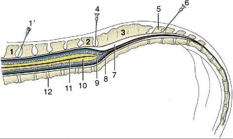
Figure 26-3 Caudal part of the bovine vertebral canal and its contents, schematic. Epidural injection sites are indicated by the needles. 1, First lumbar vertebra; 1', needle in position for flank anesthesia; 2, last lumbar vertebra (L6); 3, sacrum; 4, needle in lumbosacral space; 5, first caudal vertebra; 6, needle between first and second caudal vertebrae (tail block); 7, epidural space; 8, dura mater; 9, subarachnoid space; 10, spinal cord; 11, central canal; 12, intervertebral disk.
No cranial nuchal bursa exists, but a supraspinous bursa frequently is present between the ligament and the first few thoracic spinous processes.