THE VERTEBRAL COLUMN (See also pp. 35-38.)
The dog has 7 cervical, 13 thoracic, 7 lumbar, 3 sacral, and about 20 caudal vertebrae as a rule (Figure 12-1); the most common variation is the reduction to six of the lumbar vertebrae.
The precaudal vertebrae formula is the same in cats, in which the individual bones are generally more slender and differ from those of the dog in subtle ways that are easy to recognize but difficult to define (Figure 12-2).The intervertebral disks of both dog and cat are relatively thicker than in most species and contribute some 15% and 17% to 20%, respectively, of the total length of the column. Longitudinal growth of the column continues until approximately 12 months of age, when the epiphyses fuse with the bodies of the vertebrae—except in the sacral region where there is some delay. Table 12-1 records the ages at which the secondary ossification centers of the vertebrae appear and those at which they later fuse.

Figure 12-1 The skeleton of the dog. 1, Wing of atlas, first cervical vertebra (C1); 2, spine of axis (C2); 3, ligamentum nuchae; 4, scapula; 5, last cervical vertebra (C7); 6, cranial end (manubrium) of sternum; 7, humerus; 8, ulna; 8', olecranon; 9, radius; 10, carpal bones; 11, metacarpal bones; 12, proximal, middle, and distal phalanges; 13, sacrum; 14, hip bone (os coxae); 15, femur; 16, patella; 17, fibula; 18, tibia; 19, tarsal bones; 19', calcanean tuber; 20, metatarsal bones; T1, L1, and Cd1, first thoracic, lumbar, and caudal (tail) vertebrae.
The contours of the vertebral column do not reproduce the dorsal profile of the standing animal. The convex nape is followed by a relatively straight cervical section. A pronounced but concealed change in direction at the cervicothoracic junction redirects the column on an ascending course in relation to the contour of the back.
The caudal thoracic and lumbar segments are fairly straight (depending on the breed), but over the pelvis the column curves ventrally into the tail.The caudal end of the cervical segment is the most flexible part, and this enables the dog to reach almost every part of its trunk and limbs with its mouth. Ventral flexion to lower the head to the ground is mainly the result of movement in the cranial thoracic joints, and the cervical vertebrae are merely brought into line. Considerable mobility of the caudal thoracic and lumbar joints is necessary for the alternating sagittal flexion and extension of the back in the bounding gallop used by both cats and dogs when moving at speed. This enables the hindlimbs to be placed alongside (if not ahead of) the forelimbs, after which the hindlimb joints and those of the column extend to hurl the body forward. Lateral flexion of the joints of the thoracic and lumbar segments is surprisingly free and enables dogs to curl up when sleeping. The spine of the cat is even more supple.
At three locations in the vertebral column the dorsal parts of the vertebral arches are less closely connected and leave relatively wide interarcuate spaces: the atlan- tooccipital space between the occipital bone and the first vertebra, the atlantoaxial space between the first and second vertebrae, and the lumbosacral space between the last lumbar vertebra and the sacrum. These interarcuate spaces are of clinical importance because they can be used to allow entry to the vertebral canal for injections or for obtaining samples of cerebrospinal fluid. From the clinical point of view it is important to
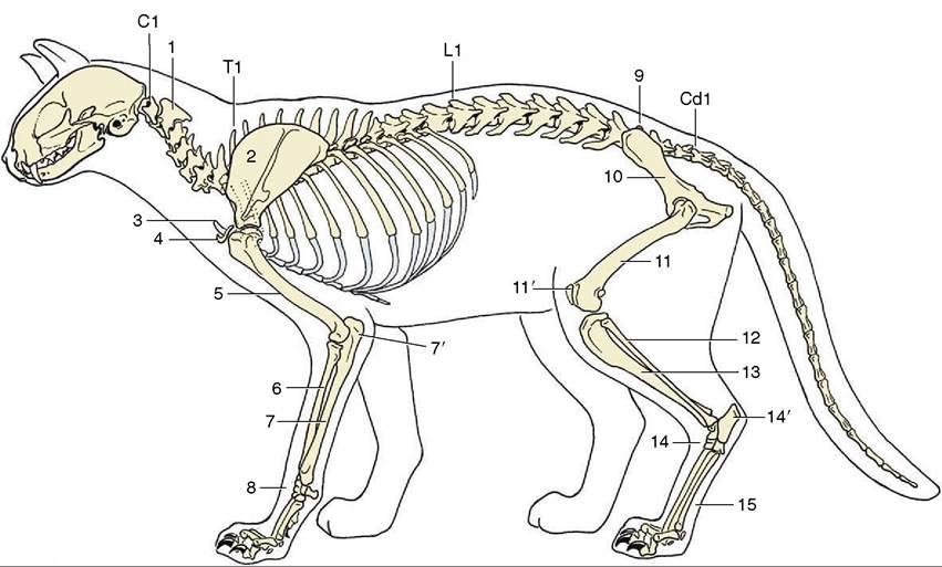
Figure 12-2 The feline skeleton. 1, Axis (C2); 2, scapula; 3, manubrium of sternum; 4, clavicle; 5, humerus; 6, radius; 7, ulna; 7', olecranon; 8, carpal bones; 9, sacrum; 10, hip bone (os coxae); 11, femur; 11', patella; 12, fibula; 13, tibia; 14, tarsal bones; 14', calcaneus; 15, metatarsal bones; C1, T1, L1, and Cd1, first cervical, thoracic, lumbar, and caudal (tail) vertebrae.
be familiar with the appearance of the vertebral column in radiographs of both juvenile and mature animals, especially at these three junctions (Figures 12-3, 12-5, and 12-6).
Because of the frequency with which spinal problems are encountered in clinical practice, it may be useful to recapitulate and amplify the descriptions given in Chapter 2.