Theovary
The paired structures known as mammalian ovaries are located within the abdominal cavity (Figure 21.2). The animal’s age, species, and reproductive state all affect the size and form of its ovaries.
However, the ovaries of monotocous animals are ovoid, while those of polytocous animals typically resemble a cluster of grapes. The ovulation fossa, a prominent depression in the kidney-shaped ovary that is the site of every ovulation, is present in mares. The cells known as surface (germinal) epithelium, which encircle the ovary and pierce through the tunica albuginea, provide granulosa cells - not eggs - to the developing ovarian follicles. The ovarian cortex, which displays corpora lutea and ovarian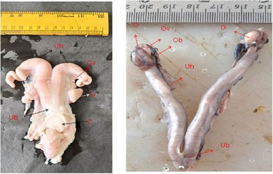
FIGURE 21.1 Female reproductive organs in goat (left) and dog (right): Ovary (O), Oviduct (Ov), Ovarian bursa (Ob), Uterine horn (Uh), Uterine body (Ub), and Cervix (C).
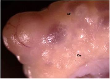
FIGURE 21.2 Gross morphology of ovary in cats: Surface follicle (SF) and Corpus albicans (CA).
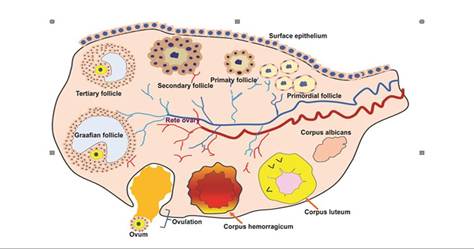
FIGURE 21.3 Schematic diagram of the ovary showing various stages of follicles and corpus luteum.
follicles at various developmental stages, is located next to the tunica (Figure 21.3). The ovarian medulla has a vast nerve and vascular supply as well as irregular fibroblastic tissue. Although they are found in the medulla as well as the cortex, smooth muscle cells are more prevalent in the perifollicular areas of the cortex. These adrenergic receptors are found in smooth muscles. The ovary’s supply of adrenaline is thought to play a role in the ovarian follicles’ selection for maturation and/or ovulation. The luteal phase is when arterial blood flow to the ovary is greatest, and ovulation is when it is lowest. Estrogen is transported from the ipsilateral ovary to the lymph vessels that drain the uterus.
21.3