THE OVARIAN FOLLICLES
The ovary serves two major purposes: (i) gametogenesis, or the production of eggs, and (ii) steroidogenesis, or the production of female sex hormones. Both are the duties of the ovarian follicle.
It is possible to categorize ovarian follicles into five different groups: primordial, primary, secondary, tertiary, and Graafian (Figure 21.3).Mesonephric cells enter the undifferentiated gonad during fetal development, undergo numerous mitotic divisions, and ultimately give rise to primordial follicles and oocytes. In postnatal life, these primordial follicles containing the primary oocytes do not go through mitotic division, in contrast to the spermatogonia in males. The thick layer of primordial follicles, which are spherical or circular in shape, lies beneath the tunica albuginea (Figure 21.4). They display oocytes with a single layer of flattened or cuboidal follicular cells surrounding them, devoid of the zona pellucida and vitelline membrane. The basement membrane is present in these primordial follicles in its entirety. There is no distinct vasculature present in these unilaminar follicles. Primordial follicles are created at birth and, as an animal ages, their quantity decreases rather than increases. A heifer calf’s ovary may contain up to 150,000 of these primordial follicles. All mammals experience this same phenomenon, though the exact number varies by species. Cats have characteristic clustering of primordial follicles, which are called Multi-oocyte follicles (Figure 21.4).
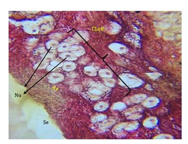
FIGURE 21.4 Photo micrograph showing clusters of primordial follicles in cats: Surface epithelium (Se), Tunica albuginea (Ta), Nuclei (Nu) & Clusters of follicles (CLoF).
A tiny portion of all follicles in a species develop into Graafian follicles and eventually ovulate.
These are known as primordial follicles. Before reaching ovulatory size, a number of them begin to grow but then begin to regress at different times. Others, however, never start the process of growth! A follicle that is growing is one that has begun to grow, has more than one layer of follicular cells, a clear zona pellucida, an oocyte with a greater diameter, and other characteristics. The follicular cells in the secondary follicles multiply and layer more, taking on polyhedral forms. These cells are now known as granulosa cells because their cytoplasm starts to show granular inclusions. Within these developing/secondary follicles, spindle-shaped cells arrange themselves outside the basement membrane to form the theca. The follicle itself enlarges, becomes more oval, and migrates from the cortex in the direction of the medulla. Follicle growth is not dependent on pituitary gonadotropins at this point. Gonadotropins cause the granulosa cells to secrete a fluid that builds up in the spaces between cells. The follicle gradually develops the antrum, a fluid-filled space, as thefluid continues to be secreted and accumulate. The follicle is now referred to as a vesicular, antral, or tertiary follicle. The corona radiata, a solid mass of cells, has now encircled the oocyte. Theca externa and interna, two layers of thecal cells, protrude from the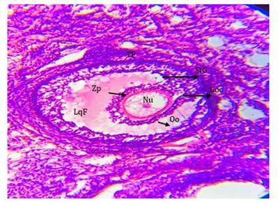
FIGURE 21.5 Photo micrograph showing Graafian follicle and cumulus oophorous oocyte complex in bitch: Stratum granulosum (StG), Cumulus oophorous oocyte complex (Coc), Tertiary follicle (TF), Oocyte (Oo), Nucleus (Nu), Zona pellucida (Zp) & Liquor folliculi (LqF).
surface of the ovary in Graafian follicles, and the oocyte’s diameter increases (varying from 80 to 120 μm in different species). The follicle wall is composed of numerous granulosa cell layers and thecal layers (Figure 21.5). Myoid cells form the follicle’s outermost wall, or theca externa, and they have smooth muscle-like properties.
Fibroblasts containing a high concentration of organelles and cytoplasmic granules form theca interna. Granulosa cells, which are isolated from thecal layers by the basement membrane, make up the innermost layer of the follicular wall. During ovulation, this membrane becomes discontinuous. Now, a solid mass of cells known as the corona radiata envelops the oocyte. The ovary’s surface is adorned with Graafian follicles, which have two layers of thecal cells called theca externa and interna. The diameter of the oocyte also grows, ranging from 80 to 120 μm depending on the species. The wall of the follicle is made up of many granulosa cell layers and thecal layers. Myoid cells, which have properties similar to smooth muscles, form the follicle’s outermost wall, or theca externa. Fibrocytes that are abundant in organelles and cytoplasmic granules form theca interna. Granulosa cells form the innermost layer of the follicular wall, which is divided from thecal layers by the basement membrane. At the time of ovulation, this membrane becomes irregular.Intrafollicular fluid pressure causes the follicle to become extremely turgid and causes the volume of the follicular fluid to increase dramatically. The granulosa cells are forced up against the follicular wall by this increase in follicular fluid, and as a result, they become known as membrana granulosa. The ovum separates from its attachment to the follicular wall and floats freely in the liquor folliculi as a result of additional follicular fluid accumulation.
21.4
More on the topic THE OVARIAN FOLLICLES:
- Abnormalities of the Ovaries
- The Doe
- REFERENCES
- Female Reproductive Disorders
- FEMALE MARSUPIALS
- Cyclic Irregularity
- Cutaneous Lymphosarcoma
- REFERENCES
- Anestrus
- Periparturient Problems