There Are Several Levels of Organization in Any Skeletal Muscle
Figure 6-2 illustrates the levels of organization in a typical skeletal muscle. Each muscle belly seen,during dissection is made up of differing numbers of muscle cells (usually called muscle fibers) that span the several inches between the origin and insertion tendons.
Γhe fibers range between 5 and 100 μm in diameter and contain multiple nuclei, multiple mitochondria, and other intracellular organelles. The outer limiting membrane of the fiber is called the sαrcolemmα. It consists of a true cell membrane, called the plasma membrane, and an outer polysaccharide layer that attaches to the tendons at the cells' extremities. Each muscle fiber is innervated by only one motor neuron, with the neuromuscular junction region located near the middle of the fiber, relative to the ends.Each muscle fiber is made up of successively smaller subunits (see Figure 6-2). Each fiber contains several hundred to several thousand myofibrils arranged in parallel along its length, like a handful of spaghetti. Each myofibril is made up of a linear series of repeating sarcomeres, the basic contractile units of the muscle fiber, which can number in the tens of thousands.
The sarcomere has a disk at each end called the Z disk. The sarcomere contains four types of large protein molecules responsible for muscular contraction, three of which are polymerized. Numerous thin protein filaments, called actin, are attached to the Z disks and extend toward the center of the sarcomere, similar to parallel fingers pointing at each other. Each actin filament consists of two intertwined, helical strands of actin protein and two such strands of tropomyosin protein, all wound together as a larger helical complex (see Chapter 1 and Figure 1-5). Also located intermittently along the tropomyosin- act in strand are complex globular protein molecules called troponin that can bind tropomyosin and actin and that have an affinity for calcium (Ca2+) ions. Suspended between and parallel to the actin thin filaments are thicker filaments of myosin protein polymers.
A myosin molecule contains a tail of intertwined helices and two globular heads that can bind both adenosine triphosphate (AFP) and actin (see Figures 1 -3 and 1 -4). Approximately 500 myosin heads of a thick myosin filament form crossbridges that interact with actin to shorten the sarcomere as the myosin heads flex and relax.Beneath the plasma membrane of the muscle cell lies the sarcoplasmic reticulum, an intracellular storage organelle that forms a reticulated network around the myofibrils (Figure 6-3). This extensive storage sac sequesters Ca2' ions in relaxed muscle and is analogous to the smooth endoplasmic reticulum in other cells.
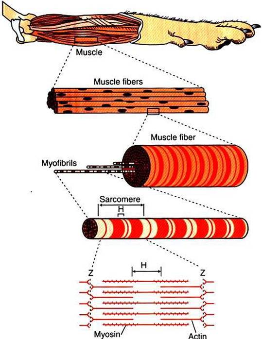
FIGURE 6-2 A typical skeletal muscle has several levels of organization. Hand Zare letters assigned to stripes seen during microscopic examination of skeletal muscle.
Located perpendicular to the long axis of the muscle fiber are tubes of plasma membrane formed by periodic invaginations of the sarcolemma. These transverse tubules, or T tubules, traverse the diameter of the muscle fiber, similar to a flexible drinking straw passing perpendicularly through the handful of spaghetti (myofibrils) noted earlier. The T tubules snake around the myofibrils, forming junctions with the network of sarcoplasmic reticulum that surrounds the myofibrils (Figure 6-4). These tubules are filled with extracellular fluid and are important because they allow the electrically excitable plasma membrane of the muscle fiber to carry the depolarization of the action potential to the interior of the fiber.