TISSUES
1. Differentiate among cells, tissues, organs, and systems as units of structure in the body.
2. Name the four basic tissues in the body.
3. Where are the general locations of epithelium?
4.
What is the function of a basement membrane?5. How does epithelium receive nutrition and discharge waste?
6. How is epithelium classified according to the number of cell layers?
7. How is epithelium classified according to the shape of the surface cells?
8. Where are the locations of endothelium, mesothelium, and mesenchymal epithelium that are derived from mesoderm and have the appearance of simple squamous epithelium that is derived from ectoderm or endoderm?
9. Know where each of the several classifications of epithelium is located.
0. What is the distinguishing feature between endocrine and exocrine glands?
11. Differentiate among holocrine, merocrine, and apocrine glands.
2. What are the two types of epithelial membranes and where are they located?
3. What are the chief functions performed by the connective tissue types?
4. What cells produce the intercellular substance of ordinary connective tissue?
L5. What are the intercellular substances of loose connective tissue? How do they differ?
6. Differentiate between dense regular and irregular connective tissue.
L7. Recognize that cartilage, bone, and blood are other elements of connective tissue.
In considering units of structure within the body, a first consideration involved the cell. The next involves tissues, which, as a unit, are composed of cells having similar features of structure and function. Two or more tissues, when combined to perform certain functions, are known as organs (e.g., the heart and liver are organs). Combinations of organs of similar or related functions, working together as a unit, are represented by body systems (e.g., the digestive system and the respiratory system).
Most of this book is organized by systems, wherein the cells, tissues, and organs for a system will be studied to recognize the contribution of each in providing for the system’s function.There are four basic tissues in the body, namely: (1) epithelial tissue (epithelium), (2) connective tissue, (3) nervous tissue, and (4) muscle tissue. Unlike nervous and muscle tissues, epithelial and connective tissues are not considered in individual chapters as a system. Therefore, some identifying features will now be given.
Epithelium
Epithelial tissues cover the body surface, line body cavities, and form glands and other structures (e.g., hair, hooves, and horns). With few exceptions, epithelium originates from ectoderm or endoderm, and the cells lie on a noncellular basement membrane. The basement membrane serves an adhesive function so that the cells are held closely to the underlying connective tissue, thereby giving greater strength to the tissue.
Epithelial tissues are not penetrated by blood vessels but rather receive nutrition and discharge waste by diffusion via blood vessels in the underlying or neighboring connective tissue.
Classification
When classified according to the number of layers of cells in the tissue, simple epithelium (one layer) and stratified epithelium (two or more layers) are recognized. There is also a classification according to shape of the surface cells, namely: (1) squamous (thin and plate-like), (2) cuboidal, being about equal in height and width (appear square in a cut perpendicular to the surface), and (3) columnar, in which the cells are taller than they are wide and in a perpendicular section are rectangular.
The types of epithelium that commonly exist throughout the body are illustrated in Figure 1-10. It will be noted that each is identified according to the number of layers and also the shape of the cell, and the following are identified:
1. Simple squamous epithelium (Figure 1-10A).
Simple squamous epithelium consists of a single layer of thin, flat cells of irregular outlines that fit together, with cement substances between their borders, to form a continuous, thin membrane.
It is not adapted to withstanding wear and tear but rather to performing a filtering function (e.g., some portions of kidney tubules).There are three tissues that have the same appearance as simple squamous epithelium but differ because they are derived from mesoderm rather than ectoderm or endoderm. In these instances they are known as endothelium, mesothelium, and mesenchymal epithelium. Endothelium is the simple layer of squamous cells forming the inner lining of the heart, blood vessels, and lymph vessels. Mesothelium is the simple squamous epithelium that lines the great body cavities (pleura and peritoneum). Mesenchymal epithelium is found in more discrete locations such as the linings of the subarachnoid spaces (in the brain) and chambers of the eye.
2. Simple cuboidal epithelium (Figure 1-10B).
This is a widely distributed tissue, and examples are found in the choroid plexus of the nervous system, the outer covering of the nervous system, the outer covering of the ovary (reproductive system), and the lining of follicles in the thyroid gland (endocrine system).
3. Simple columnar epithelium (Figure 1-10C).
This tissue provides the lining for the digestive tract. The cells may be absorptive, secretory, or both. A common secretory function of these cells is secretion of mucus on the surface of epithelial membranes, and in this capacity they provide a protective function. There are also simple columnar ciliated tissues. Cilia are motile extensions of a cell surface that move tubular contents in a single direction. An example of their presence is in the uterine tubes (oviducts).
4. Transitional epithelium (Figure 1-10D).
This tissue is common to the lining of the muscular urinary bladder. It is a stratified epithelium with a varied appearance depending on the fill of the bladder. When the bladder contracts, the epithelium piles up into many layers, but when the bladder fills and is stretched, only two or three layers of cells can be seen.
5.
Pseudostratified ciliated columnar with goblet cells (Figure 1-10E).These tissues seem to consist of many layers but actually have only one layer. The one shown is ciliated, but there are also those that are nonciliated. The stratified appearance is caused by some of the cells being short and other taller cells overlapping them. They both share a common basement membrane. The type shown, with cilia and goblet cells, are found in the respiratory tract. The goblet cells provide for a wet surface for entrapment of inhaled particles, and the cilia direct the wet surface toward the mouth.
6. Stratified squamous (Figure 1-10F and G).
Stratified membranes serve chiefly to protect. They can withstand more wear and tear than simple membranes. There are different kinds and degrees of protection needed at different places in the body and, accordingly, stratified membranes have dissimilarities. The kind shown in the illustration is nonkeratinized stratified squamous epithelium (Figure 1-10F) and is found on wet surfaces subjected to wear and tear. The inside of the mouth and esophagus have this lining, giving protection from coarse foods. Only the surface cells are actually squamous, the deepest layer (on the basement membrane) of cells is columnar. As the deep layer cells undergo mitotic division, the outer cells flatten, die, and finally slough (separate) from the surface. In this way the tissue renews itself. The epidermis (outer layer) of skin is stratified squamous keratinized tissue (Figure 1-10G). This differs from nonkeratinized epithelium in that the superficial cells are keratinized (also called cornified). The cells of this type are also fused with each other. The cornified and fused layer minimizes fluid loss from the body by evaporation and gives greater protection from wear and tear.
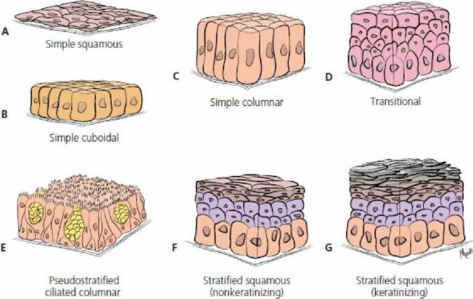
■ FIGURE 1-10 Epithelial tissue classifications. The epithelial cells are shown lying on a noncellular basement membrane that serves an adhesive function holding the cells closely to the underlying connective tissue.
Glands
The glands of the body are classified as exocrine or endocrine. Both are secretory, but exocrine glands are those that have secretions to the outside of the body (this includes organ lumens) and endocrine glands are those that secrete within the body. Exocrine glands must be provided with ducts, which are tubes that convey the glandular secretions to a free surface of the body. Because endocrine secretions are those within the body, no ducts are needed and so they are often referred to as ductless glands.
Development of both glands is shown schematically in Figure 1-11. It is noted that both originate as a result of surface epithelial cells growing, in the form of either a cord or a tubule, into the connective tissue beneath the membrane. After invasion of the connective tissue, a gland is formed by means of further proliferation and differentiation. The epithelial connection between the gland and surface is retained for exocrine glands, whereas the connection disappears for endocrine glands. Those cells that form the secretory unit secrete their substances into a central cavity or lumen.
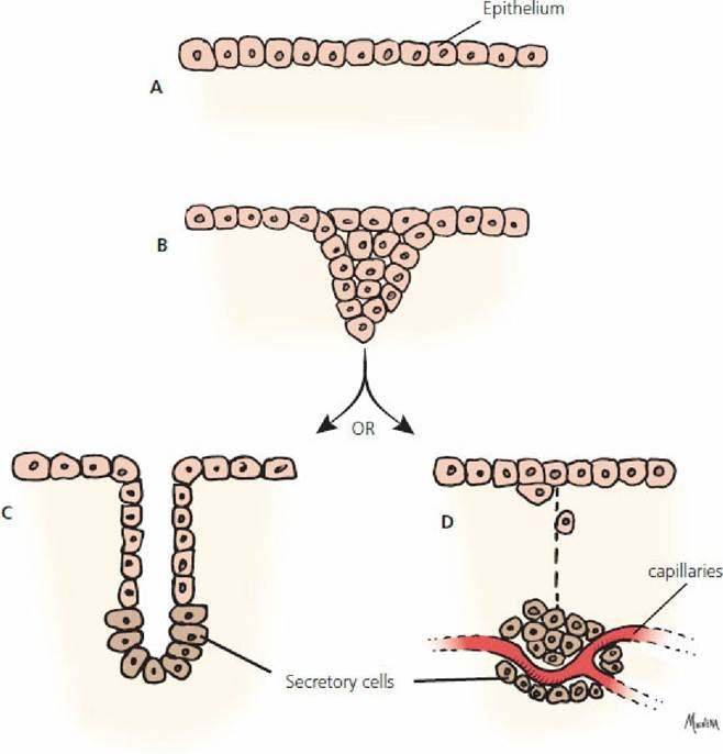
■ FIGURE 1-11 The development of exocrine and endocrine glands. A. Surface epithelial cells. B. Epithelial cells invading into the connective tissue. C. An epithelial connection is maintained in exocrine glands but is lost for endocrine glands (D).
Holocrine, merocrine, and apocrine glands refer to the manner in which the secretory cells of the gland elaborate their secretions. A cell within holocrine glands accumulates secretory products in its cytoplasm and then dies and disintegrates. The dead cell and its products constitute the secretion (i.e., the entire cell is secreted). The sebaceous (oily, fatty) glands of the skin are of this type.
Merocrine glands secrete without any part of the cell being lost. Secretory granules are cytoplasmic inclusions and, although produced by the cytoplasm, they are not actually part of the cytoplasm.
Therefore, the secretory granules pass into the lumen of the secretory unit without loss of the secretory cells’ cytoplasm. The pancreas and salivary glands are in this group.Apocrine glands are intermediate between holocrine and merocrine glands because their secretions gather at the outer ends of the gland cells and then pinch off to form the secretions. The mammary glands and some sweat glands belong to this group.
Epithelial Membranes
Epithelial membranes consist of a surface layer of epithelium and an underlying layer of connective tissue. Two kinds that are of importance in the body are mucous membranes and serous membranes.
Mucous membranes, referred to as mucosae, line the hollow organs and cavities that open on the skin surface of the body. These membranes line most of the organs of the digestive, respiratory, urinary, and reproductive systems. The surface epithelium may vary in type, but it is always kept moist by mucus. The connective tissue underlying the epithelium is referred to as the lamina propria.
Serous membranes, referred to as serosae, line the body cavities and cover the surfaces of related organs. The surface epithelium is mesothelium over a thin layer of loose connective tissue. The mesothelium provides fluid that serves to moisten and lubricate. Pleura (lining the thorax),
pericardium (lining the cavity around the heart), and peritoneum (lining the abdomen and pelvic cavities) are examples of serous membranes.
Connective Tissue
A wide range of tissues that share a common origin from mesoderm represents connective tissues. The chief functions performed by the various cells of the different types of connective tissue follow: (1) production of intercellular substances, (2) storing fat (adipocytes), and (3) production of the various blood cells, which in turn have specific functions (e.g., phagocytosis of bacteria and production of antibodies). The intercellular substance of chondrocytes and osteocytes (cartilage and bone) are connective tissues specialized for the support of the body. Cartilage, bone, and blood are elements of connective tissue that will be described in separate chapters.
Ordinary Connective Tissues
Ordinary connective tissues connect other tissues and are classified as either loose or dense.
Loose connective tissue contains a variety of different cell types. Loose connective tissue is widely distributed in the body, where it makes up the subcutaneous tissue or superficial fascia. It penetrates between organs to fill space and bind structures together. Because of its loose nature, it allows for movement of muscles relative to one another. Fibroblasts are the cells that produce the intercellular substance of ordinary connective tissue. When less active during adult life, fibroblasts are often referred to as fibrocytes.
Important intercellular substances of loose connective tissue are (1) collagenous or white fibers, (2) elastic or yellow fibers, (3) reticular fibers, and (4) amorphous ground substance.
Collagenous fibers appear as wavy ribbons. They are strong and inelastic and are composed of collagen, a family of closely related proteins. Elastic fibers are long cylindrical threads or flat ribbons. They tend to regain their original shape after being stretched. They are found in the walls of elastic arteries and are mixed with other tissues wherever elasticity is needed. Reticular connective tissue fibers are fine and highly branched. They make up part of the framework of endocrine and lymphatic organs and also form networks where structures are adjacent to connective tissue, as found along the blood vessels, in basement membranes, and around nerve, muscle, and fat cells. Like collagenous fibers, they are inelastic. The above fibers are imbedded in amorphous (without form) ground substance. The viscosity of amorphous ground substance varies from fluids to gel. Figure 1-12 illustrates cells and fibers that might be seen in a microscopic section of loose connective tissue.
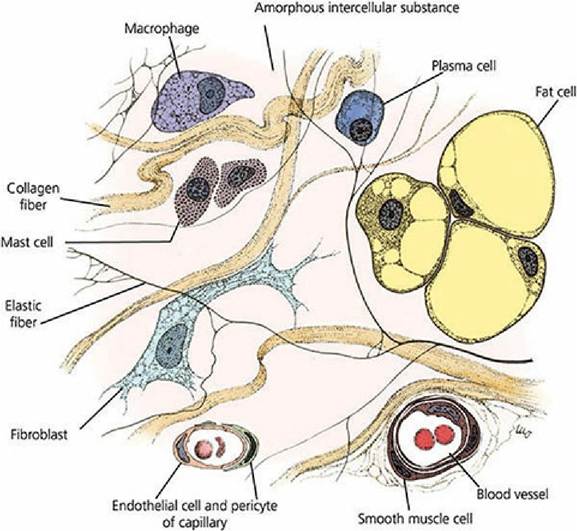
■ Figure 1-12 Fibers and cells of loose connective tissue. Mast cells are usually found close to small blood vessels and have granules containing potent inflammatory mediators (e.g., histamine). Macrophages are phagocytic and plasma cells are the source of circulating antibodies (immunoglobulins). Pericytes are intimately associated with blood capillaries and venules, providing a potential source of new fibroblasts and smooth muscle cells. (From Cormack DH. Ham’s Histology. 9th edn. Philadelphia, PA: JB Lippincott Company, 1987.)
Dense connective tissues contain essentially the same fiber elements as loose connective tissues. There are two types, dense regular and dense irregular. The regularity relates to the arrangement of the fiber elements. In dense regular connective tissue, the fibers (especially collagenous fibers) are arranged in parallel bundles forming tendons. In ligaments, the collagenous fibers are not as regularly arranged and may be mixed with elastic fibers. The ligamentum nuchae in the necks of grazing animals has a predominance of elastic fibers. In dense irregular connective tissue, the collagenous fibers are interwoven and compacted to form a dense matting. This type is found in the dermis of the skin. The dermis of the skin is used in the production of leather. It is treated with tannic acid after the epidermis is removed.
Cartilage, bone, and blood are other elements of connective tissue that will,be described separately in respective chapters.
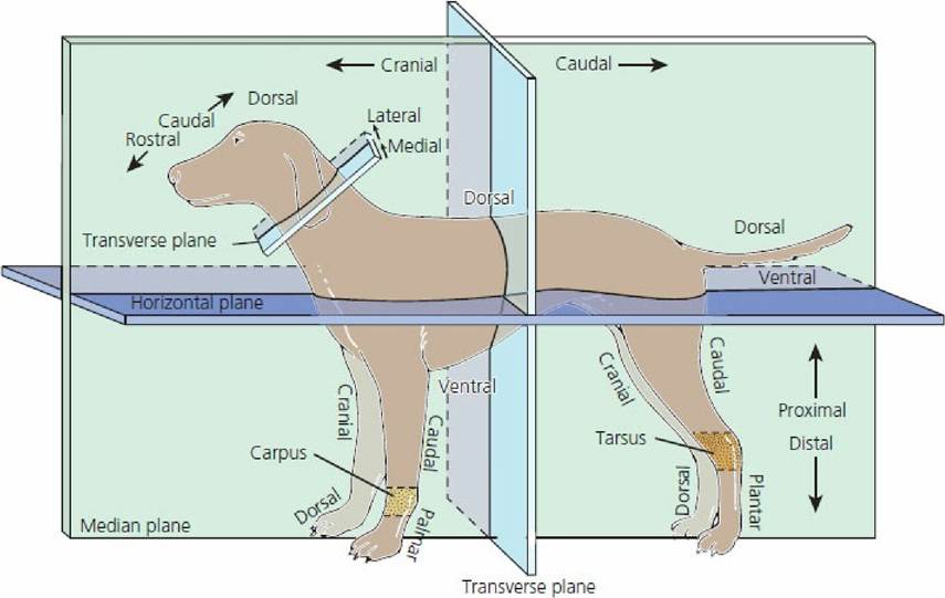
■ FIGURE 1-13 Directional terms and planes as applied to four-footed animals. The stippled areas represent the carpus and tarsus on the forelimbs and hindlimbs, respectively.
■