EMBRYOLOGY
1. Differentiate between diploid and haploid.
2. How does meiosis contrast with mitosis?
3. What is meiosis accompanied by division of the cells called in the female and in the male?
4.
Define embryology.5. Differentiate among gamete, zygote, morula, and blastocyst.
6. What does the trophoblast contribute to in fetal development?
7. Name the three germ layers established as embryonic development proceeds.
8. What two major events are signified by the development of the germ layers?
Fertilization is the first event of reproduction at the cellular level and requires the joining of the female sex cell (gamete), known as the oocyte, with the male gamete, known as the spermatozoon. So that the fertilized oocyte will have the normal number of chromosomes (diploid or 2n), each gamete must be reduced in chromosome numbers by one-half (haploid or n) while still in the reproductive systems of the respective female and male. This reduction in chromosomes is called meiosis, in contrast to mitosis, whereby each cell after division retained the 2n chromosome number. Meiosis accompanied by division of the cells is called oogenesis in the female and spermatogenesis in the male. The joined gametes, now known as a zygote, will have the proper number of chromosomes (2n) for the species, and further development beyond fertilization will proceed by mitosis. Fertilization and the beginning of mitosis for the formation of a new individual are shown in Figure 1-7. For further details of spermatogenesis, oogenesis, and fertilization, see Chapters 14 and 15.
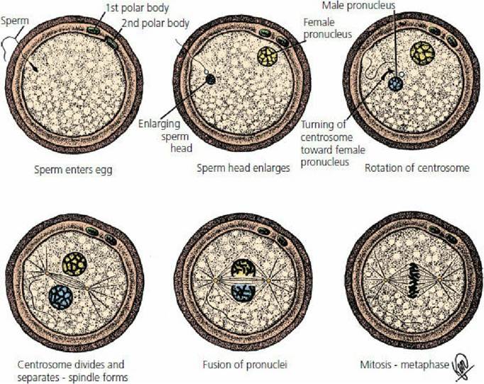
■ FIGURE 1-7 Schematic diagrams of fertilization. Meiosis in spermatozoa and oocytes (division of chromosome numbers by one-half) occurs while in respective male and female reproductive systems.
Entrance of a spermatozoon into an oocyte is followed by fusion of respective pronuclei to form a zygote with a proper chromosome number (2n or diploid). Cell division will proceed by mitosis to form a new individual. (From Crouch JE. Functional Human Anatomy. 4th edn. Philadelphia, PA: Lea & Febiger, 1985.)Embryology is the study of prenatal (before birth) development of an individual and, as indicated above, it begins with the zygote. Mitotic divisions continue and form a cluster of cells known as a morula that proceeds to a blastula (Figure 1-8). The cavity of the blastula, the blastocele, is formed when uterine fluid diffuses into the spaces between the cells of the morula. As the fluid accumulates, it gradually separates the cells into an outer layer of cells called the trophoblast and an inner cell mass that forms the body of the embryo (Figure 1-qA). The trophoblast contributes to the fetal placenta (extraembryonic membranes) that secures the position of the embryo in the uterus and provides for its nutrition from the maternal connection (see Chapter 15).
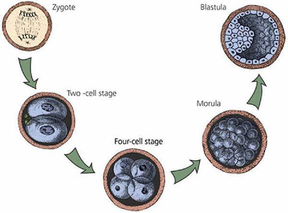
■ FIGURE 1-8 Continued mitotic division from zygote to blastula. (From Frandson RD, Wilke WL, Fails AD. Anatomy and Physiology of Farm Animals. 7th edn. Ames, IA: Wiley-Blackwell, 2009.)
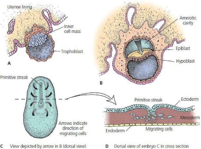
■ FIGURE 1-9 The formation of the germ layers, ectoderm, mesoderm, and endoderm. A. Embryo embeds in the wall of the uterus. B. Formation of epiblast and hypoblast layers. The amniotic cavity is formed dorsal to the epiblast, and the hypoblast cells migrate to line the cavity of the blastula (blastocele), which becomes endoderm. C. Embryo viewed from above. The primitive streak is a thickening of epiblast cells on the longitudinal axis that migrate toward the primitive streak and become ectoderm. D. Cross-section through the region of the primitive streak showing migration of cells between ectoderm and endoderm that become mesoderm.
(From Frandson RD, Wilke WL, Fails AD. Anatomy and Physiology of Farm Animals. 7th edn. Ames, IA: Wiley-Blackwell, 2009.) The portion of the inner cell mass closest to the trophoblast is the epiblast and the portion adjacent to the blastocele is the hypoblast (Figure 1-qB). The cavity formed dorsal to the epiblast is the amniotic cavity of the embryo (see Chapter 15). Proliferating hypoblast cells migrate to line the blastocele. This lining becomes the endoderm. The endoderm grows into the blastocele and generates the lungs, gut, liver, and other visceral organs. The ectoderm develops from proliferating outer cells of the inner cell mass (epiblast cells) and migrates toward a longitudinal axis location known as the primitive streak, a thickening of epiblast cells (Figure i-qC). Skin and all of its derivatives (e.g., hair, hooves, mammary glands) and the entire nervous system are formed from ectoderm. The cells between ectoderm and endoderm become mesoderm (Figure 1-qD). The mesoderm grows between the ectoderm and endoderm and splits into two layers that form a cavity between the two layers known as the coelom (precursor of body cavities). The pericardial, pleural, and abdominopelvic cavities are derived from the coelom. Skeletal, smooth, and cardiac muscle, the kidneys, the skeleton, and other connective tissues develop from mesoderm. The establishment of the germ layers is the first segregation of cell groups clearly distinct from one another by way of their definite relations within the embryo. Also, establishment of the germ layers marks the transition between that period of development when an increase in the number of cells was the only outstanding event to one when differentiation and specialization are the dominating aspects of growth. The germ layers are the source of all body structures.