Tributaries of the Caudal Vena Cava
The paired internal iliac veins join the external iliac veins, and the two common iliac veins converge at the level of the seventh lumbar vertebra in carnivores (L6 in the horse and ruminants, L6/L7 in the pig).
The caudal vena cava then continues cranially in the roof of the abdomen and on the right side of the aorta. When the vena cava reaches the liver, it inclines ventrally before passing through the diaphragm at the caval foramen (see Figure 8.3).9.1.1 Deep circumflex iliac veins
These paired veins join the vena cava at the level of the junction of the common iliac veins. They correspond to the arteries of the same name.
9.1.2 Lumbar veins
Seven pairs of lumbar veins correspond to the lumbar segmental arteries. The first two lumbar veins (i.e. the most caudal) are tributaries of the azygos vein. In the horse and carnivores it is the right azygos vein that becomes functional. In the ox, sheep and pig it is the left azygos vein that persists. The right azygos vein (formerly called the hemiazygos vein) drains into the final part of the cranial vena cava (or sometimes the right atrium of the heart). The left azygos vein enters the right atrium at the coronary sinus, the terminus of the great coronary vein. The azygos vein drains most of the blood from the vertebral venous sinuses.
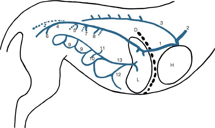
Figure 9.1 Diagram of the abdominal veins of the dog. Note that the right veins only are labelled where the veins are paired. D = diaphragm; L = liver; H = heart. 1 = caudal vena cava;
2 = cranial vena cava; 3 = right azygos; 4 = external iliac; 5 = right deep circumflex; 6 = right internal iliac; 7 = right testicular/ovarian; 8 = right renal; 9 = jejunal; 10 = caudal mesenteric; 11 = splenic;
12 = gastroduodenal; 13 = portal
9.1.3 The gonadal veins
The testicular veins receive blood from the testis and epididymis. They become coiled, tortuous and intertwined with the artery, lymphatics and nerves; this arrangement is called the pampiniform plexus (see Sections 16.4 and 16.5).
The right testicular vein enters the caudal vena cava just caudal to the right renal vein. The left testicular vein or the left ovarian vein enters the left renal vein.The right ovarian vein is at the same location as the male vessel, but it is less tortuous than its homologue. It receives several tributaries from the ovary and adjacent tissues. The uterine arterial tributary forms an anastomosis with the uterine vein (a tributary of the external iliac vein) within the broad ligament.
9.1.4 Renal veins
The paired renal veins are quite short, and they are 6-8 mm in diameter. The left renal vein receives the left testicular vein in the male and the left ovarian vein in the female.
9.1.5 Phrenicoabdominal veins
These paired veins enter the vena cava just cranial to the renal veins. They cross the ventral surface in a groove on the ventral surface of the corresponding adrenal gland (suprarenal gland, NAV). They receive blood from the diaphragm and the body wall.
9.1.6 Hepatic veins
There are at least 20 tributaries of various sizes draining the liver lobes to the vena cava before it passes through the diaphragm.
9.1.7 Phrenic veins
These paired veins drain the diaphragm and enter the vena cava just as it passes through the diaphragm.
9.2