Upper Respiratory Tract
Nose
The nose of domestic animals comprises the parts of the face rostral to the eyes and dorsal to the mouth. The external nares (nostrils) are the external openings of the respiratory tract (Fig.
19-1). Their size and shape, highly variable among domestic farm animals, are largely dictated by the nasal cartilages that form this most rostral end of the respiratory tract. in addition to these hyaline cartilages, the pig also possesses a rostral bone in the tip of its flat, disklike nose. This is presumably an adaptation to the rooting habits of the pig.The lateral aspect of the nose is covered with typical hairy skin, which contains both sebaceous and sweat glands. The hairless region of the most rostral parts of the nose in species other than the horse contains no sebaceous glands but does have numerous sweat glands, which keep the region around the nostrils moist. This area is the planum nasale in the sheep and goat, planum rostrale in the pig, and planum nasolabiale in the cow. The grooves and bumps in the plana are distinctive enough to allow nose prints to be used for positive individual identification.
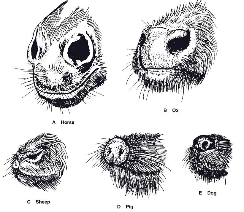
Figure 19-1. External nares of various species. The horse (A) lacks a planum, its nose being instead covered with fine hair. The ox (B) possesses a nasolabial planum, and the small ruminants (C) and dogs (E) have a nasal planum. The pig's external nose (D) features a rostral planum that is supported by a rostral bone.
The equine nose lacks a planum, being instead covered with short, fine hairs. The lateral wall of the external naris is flexible, allowing for an enormous range of diameters. During exertion, the lateral wall is dilated, creating a wider, lower-resistance passageway for the movement of air. in this, the nostril is aided by the presence of a short blind-ended diverticulum lateral to the true nasal cavity.
This “false nostril” (nasal diverticulum) is probably a construct that aids in passive dilation of the nostrils during vigorous ventilation.The nasal cavity is separated from the mouth by the hard and soft palates and separated into two isolated halves by a median nasal septum. The rostral part of the septum is cartilaginous, whereas the most caudal part is created in part by a plate of bone. Each half of the nasal cavity communicates with the nostril of the same side rostrally and with the pharynx caudally by way of the choanae (caudal nares).
The nasal cavity is lined with mucous membrane that covers a number of scroll-like conchae (turbinate bones) arising from the bones of the lateral wall (Fig. 19-2). The two major conchae (dorsal concha and ventral concha) occupy rostral parts of the nasal cavity; caudal parts are filled with ethmoidal conchae. The vascular mucous membrane covering these conchae helps to warm and humidify inspired air. The mucous membrane investing the ethmoidal conchae is the olfactory epithelium. It contains the sensory endings of the olfactory nerve (cranial nerve I), which mediates the sense of smell (see Chapter 11).
The actual air space of each half of the nasal cavity is divided by the conchae into
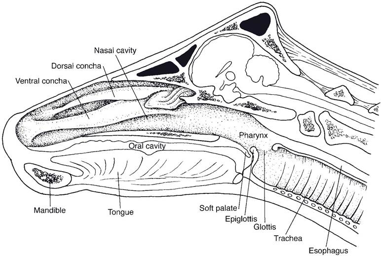
Figure 19-2. Median section of the bovine head with nasal septum removed. (Reprinted with permission of Wiley-Blackwell from Reece W.R. Physiology of Domestic Animals. 2nd ed. Baltimore: Williams & Wilkins, 1997.)
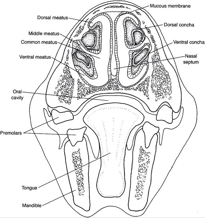
Figure 19-3. Transverse section through the nasal cavity of the horse, illustrating meatuses created by conchae. (Reprinted with permission of Wiley-Blackwell from Reece W.R. Physiology of Domestic Animals. 2nd ed. Baltimore: Williams & Wilkins, 1997.)
longitudinally arrayed nasal meatuses (Fig. 193). The dorsal nasal meatus is between the dorsal concha and the roof of the nasal cavity.
The middle nasal meatus is between the two conchae. The ventral nasal meatus is between the ventral concha and the floor of the nasal cavity. The common nasal meatus communicates between the others and is adjacent to the nasal septum. A nasogastric tube introduced through the nostrils into the nasal cavity and advanced into the esophagus for administration of medications is directed into the space that is the junction of the common and ventral nasal meatus.Paranasal Sinuses
Many of the cranial bones contain air-filled cavities, paranasal sinuses, that communicate with the nasal cavity. These sinuses probably provide some protection and insulation to the head. Although there are some species differences, all farm animals have maxillary, frontal, sphenoidal, and palatine sinuses in the bones of the same name (Fig. 19-4). In horses, several uppercheek teeth project into the maxillary sinus, which may become infected by diseased teeth (see Figure 20-3). The frontal sinus is especially capacious. in horned cattle, an extension of the
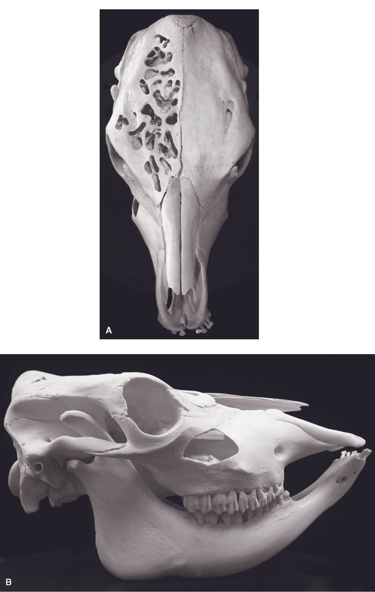
Figure 19-4. Paranasal sinuses of the ox demonstrated by sculpting of bones. A) Frontal sinus. B) Maxillary sinus.
frontal sinus, the cornual diverticulum, extends well into the bony core of the horn (Fig. 13-7). Dehorning of animals older than 3 or 4 months of age usually exposes the interior of the frontal sinus and predisposes it to infection.
Pharynx
The pharynx is a common soft tissue conduit for food and air, lying caudal to the oral and nasal cavities. openings into the pharynx include the two caudal nares (choanae), two auditory tubes from the middle ears, the oral cavity, the larynx, and the esophagus. The walls of the pharynx are supported by striated muscles whose actions assist in deglutition (swallowing) and phonation. in normal circumstances, foodstuffs and liquids are directed into the esophagus from the mouth (and vice versa during rumination) with a coordinated movement of pharyngeal and laryngeal muscles that excludes these substances from the airways.
Larynx
The larynx is the gatekeeper to the entrance of the trachea. it maintains a rigid, boxlike shape via a series of paired and unpaired cartilages that are moved relative to one another by striated laryngeal muscles. The larynx’s primary function is to regulate the size of the airway and to protect it by closing to prevent substances other than air from entering the trachea. it is capable of increasing the diameter of the air passageway during forced inspiration (as during heavy exercise) and closing the opening during swallowing or as a protective mechanism to exclude foreign objects. Secondarily, the larynx is the organ of phonation (vocalization), hence its common name, voice box. Contraction of muscles in the larynx changes the tension on ligaments that vibrate as air is drawn past them; this vibration produces the voice.
Five mucous membrane-lined cartilages make up the larynx in most domestic animals (Fig. 19-5). The unpaired, spade-shaped epiglottic cartilage (epiglottis), which lies just caudal to the base of the tongue, is mostly elastic cartilage. During deglutition, movements of the tongue and larynx fold the epiglottis caudad so that it covers the entrance into the larynx.
The thyroid cartilage resembles a taco shell, consisting of two parallel plates, the laminae, on each side, joined on the ventral midline. in the human, the thyroid cartilage, or Adam’s apple, projects from the ventral aspect of the neck. The laminae of the thyroid cartilage have processes that articulate with other cartilages and give attachment to a number of muscles.
The cricoid cartilage is shaped like a signet ring, with the broad portion dorsal. The cricoid cartilage articulates with the thyroid cartilage and the two arytenoid cartilages (discussed later) cranially and attaches to the first cartilaginous ring of the trachea caudally.
The paired arytenoid cartilages are irregular in shape, with processes that vary between species. The arytenoid cartilages of all species have a ventral vocal process, to which is attached the vocal ligament (vocal cord).
The vocal ligaments mark the division between the vestibule, the laryngeal space cranial to them, and the infraglottic cavity, the space caudal to them (Fig. 19-5). The slitlike gap between the two vocal ligaments is the rima glottidis. Movement of the arytenoid cartilages changes the tension and angle of the vocal ligaments, closing the glottis or adjusting the pitch of the voice as the ligaments vibrate.Horses and swine also possess a vestibular (ventricular) ligament cranial to the vocal ligament. An outpocketing of mucous membrane between the two ligaments forms a blind pouch called the lateral ventricle.
The vagus nerve (cranial nerve X) carries fibers that innervate the muscles (motor) and the mucous membranes (sensory) of the larynx. Most of the muscles receive their motor innervation from the recurrent laryngeal nerve, which for embryological reasons branches from the vagus within the thorax and must return craniad along the trachea to reach the larynx. This very long course of the axons in the recurrent laryngeal nerve (from the brainstem, where the neuronal cell bodies reside, down the neck into the thorax and back up the neck to the
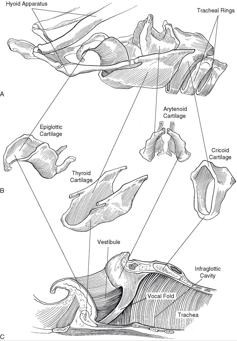
Figure 19-5. Larynx of the horse. A) lateral view of the assembled larynx, which is attached rostrally to the hyoid apparatus and caudally to the trachea. B) Individual cartilages of the equine larynx. C) Internal anatomy of the larynx.
larynx) makes them susceptible to trauma and metabolic diseases.
Injury to one of the recurrent laryngeal nerves will result in paralysis of most of the laryngeal muscles on the same side. Paralysis of the muscle that abducts the arytenoid cartilages and thereby increases the diameter of the airway (the dorsal cricoarytenoid muscle) results in a condition in horses called laryngeal hemiplegia or roaring. A horse that is a roarer cannot expand the rima glottidis during forceful inspiration and consequently has difficulty bringing sufficient air into the lungs when exercising.
The flaccid vocal ligament flutters as air moves rapidly past it, generating a loud, hoarse sound. Roaring usually is due to unilateral damage to the left recurrent laryngeal nerve, as this nerve’s course is somewhat longer than that of the right recurrent laryngeal nerve.Trachea and Bronchi
The trachea extends from the caudal end of the larynx to the bronchi (Fig. 19-6). It is formed by a series of C-shaped hyaline tracheal cartilages that provide cross-sectional rigidity to resist collapse and are joined one to another by elastic annular ligaments that permit the trachea considerable flexibility to follow movements of the neck. The dorsal side of the trachea is completed by connective tissue and the m. trachealis, a smooth muscle whose tone affects the diameter of the trachea.
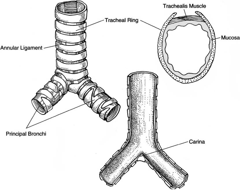
Figure 19-6. Basic anatomy of trachea and principal bronchi.
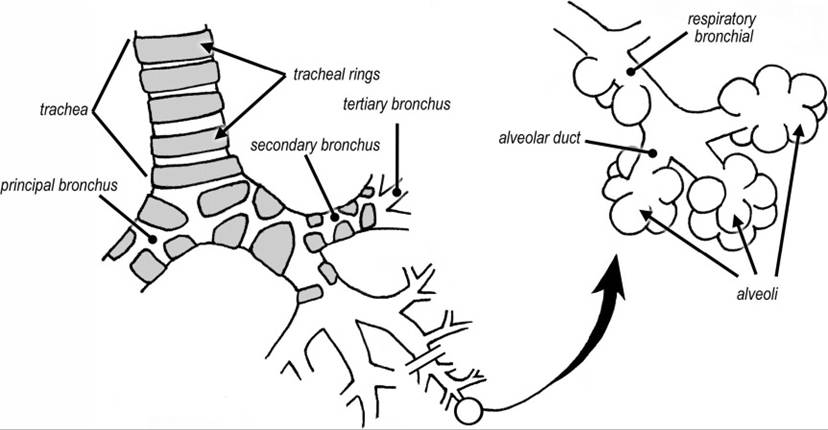
Figure 19-7. General design of airway branching from trachea to alveoli.
The trachea passes caudad as far as the base of the heart, where it divides into two principal bronchi, one for each lung (Fig. 19-6). The ruminants and pig have an additional tracheal bronchus arising cranial to the principal bronchi; it supplies the cranial lobe of the right lung. The principal bronchi branch into secondary (also called lobar) then tertiary bronchi, subsequent branches becoming smaller and smaller. The walls of these bronchi are supported by cartilaginous plates. When the airways divide to the extent that they are less than 1 mm in diameter, the cartilage disappears, and these airways are called bronchioles. The bronchiole eventually branches into several alveolar ducts, which terminate in clusters of air sacs, the alveoli. It is here that the exchange of gases with the blood takes place. Some terminal bronchioles have alveoli in their walls, hence are called respiratory bronchioles (Fig. 19-7).