Thorax
The thorax is bounded cranially by the first pair of ribs, the first thoracic vertebra, and the cranial part of the sternum. This ring of skeletal elements is the thoracic inlet.
The dorsal part of the thorax is defined by the thoracic vertebrae and epaxial muscles, and the ventral part, by the sternum. The ribs and costal cartilages, linked by intercostal muscles, create the lateral walls. The overall shape of the thorax is that of a cone with the apex at the thoracic inlet. The base of the cone is covered by the dome-shaped diaphragm.Lungs
Each lung is roughly conical, with the base resting against the cranial side of the diaphragm and the apex in or close to the thoracic inlet. The medial aspect of each lung features an indentation, the hilus, where the principal bronchus, pulmonary vessels, lymphatics, and nerves enter and leave the lung.
Lobes of the lungs are defined by the presence of lobar (secondary) bronchi. The lobes are grossly distinguishable in most species by deep fissures in the ventral part of the lung. In ruminants and the pig, the left lung is divided into cranial (apical) and caudal (diaphragmatic) lobes, with the cranial lobe having a further division into cranial and caudal parts.
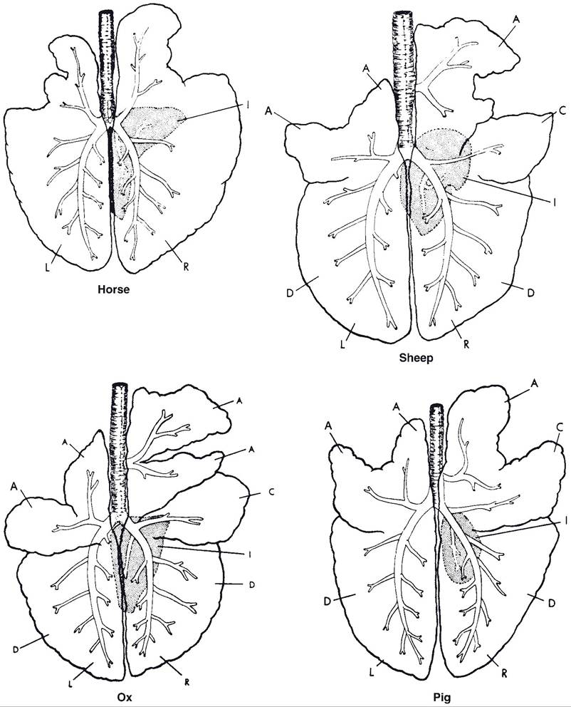
Figure 19-8. Lungs of horse, sheep, ox, and pig. L, left; R, right; A, cranial (apical); C, middle (cardiac); D, caudal (diaphragmatic); I, accessory (intermediate). (Redrawn after Nickel R., Schummer A., and Seiferle E. Lehr- buch der Anatomie der Haustiere. Berlin: Paul Parey, 1960. Permission from Wiley-Blackwell.)
The right lung in these animals is divided into four lobes: cranial and caudal lobes as on the left, plus a middle lobe between these and a ventrocaudal accessory lobe near the midline (Fig. 19-8). A more or less distinct gap between lobes along the ventral margin of the lungs is usually identifiable.
This is the cardiac notch, and it tends to be larger on the right side (Fig. 19-9). At the cardiac notch, the heart makes contact with the thoracic wall, a fact that is exploited for echocardiogram imaging (wherein the ultrasound device is applied to the thoracic wall, where no lung tissue intervenes between it and the heart) and pericardiocentesis (introducing a needle through the body wall to sample fluid from the pericardial sac).Both right and left lungs of the horse have cranial and caudal lobes, distinguished by the
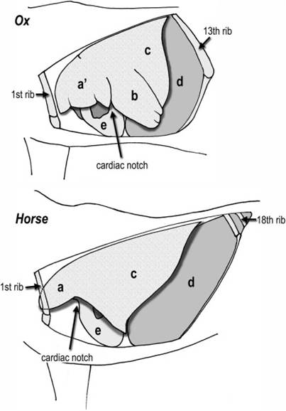
Figure 19-9. Disposition of the left lung in the ox (above) and horse (below), shown in a cutaway view of the thorax. a, cranial lobe; a', cranial part of cranial lobe (ox only); b, caudal part of cranial lobe (ox only); c, caudal lobe; d, diaphragm; e, heart.
cardiac notch. The right equine lung also has a small accessory lobe.
After an animal has taken one breath, the lungs always retain a significant volume of air, even in pathologic conditions of collapsed lung. In the fetus, however, the lungs are nearly the consistency of liver, contain no air, and sink in water. Whether the lungs will sink or float in water is a standard test to determine whether a newborn animal was born dead, in which case the lungs sink, or drew at least one breath, in which case the lungs float.
Pleura
The thoracic cavity is lined by a serosa, the pleura. The smooth surfaces of the pleura are lubricated with a scant amount of serous fluid, facilitating frictionless movement of the lungs during respirations. The pleura is attached to the bony and muscular elements of the thorax by endothoracic fascia, a thin connective tissue layer.
The pleura consists of two separate sacs, one surrounding each lung. The pleura that lines the thorax is the parietal pleura, and the pleura that covers the lungs is the visceral pleura. The pleural cavity, between parietal and visceral pleurae, is a potential space only.
This pleural cavity normally contains nothing except a small amount of serous fluid; conditions that introduce fluid or gas (e.g., pus, blood, air) into the pleural space compress and may collapse the lung associated with that space.The junction of the two pleural sacs near the midline of the thorax forms a double layer called the mediastinum in which are found the heart, great vessels, esophagus, and other midline structures. The caudal vena cava and right phrenic nerve (nerve innervating the right side of the diaphragm) are enclosed in a distinct fold of pleura, the plica venae cavae. The mediastinum of cattle is thick and forms a complete barrier between the right and left pleural cavities. in horses, parts of the mediastinum are thin, and openings between the two cavities either occur naturally or are readily created. For this reason infections or air in one pleural space may stay unilaterally contained in cattle, whereas they spread rapidly to involve both sides in the horse.