UpperAirway Obstruction in a Boston Terrier
History. A Boston terrier exhibits signs of severe respiratory distress. It has difficulty inhaling and makes a snoring sound during inhalation. The effort of walking magnifies the distress.
An arterial blood gas (ABG) sample reveals that Paeo2 is 80 mm Hg (normal, 40 mm Hg), pH is 7.3, [HCO3-] is 39 mEq∕L, and base excess is 10 mEq/L (Figure 52-6).Clinical Examination. Examination reveals excessively narrowed (stenotic) nares and excessive folds of tissue in the soft palate, the latter occluding the glottis. The larynx and trachea appear normal.
Treatment. Reconstructive surgery is performed on the dog to enlarge the nares and remove the excessive tissues from the palate. Two weeks after surgery, respiratory distress is greatly reduced. ABG analysis reveals that Paco2 is 45 mm Hg, pH is 7.39,1HCO3-] is 27 mEq/L, and base excess is 2 mEq/L.
Comment. Before surgery the animal is acidotic with an elevated Paco2 and base excess. Only the high Paeo2 explains the acidosis; therefore the dog has respiratory acidosis. The increase in [HCO3-] (normal, 24 mEq/L) is caused primarily by creation of new HCO3 (a base excess) by the kidneys and indicates the condition is of at least several days’ duration. Respiratory acidosis is caused by alveolar hypoventilation resulting from the upper airway obstruction. Surgery corrects the obstruction and alleviates the hypoventilation. This returns the pH to a more normal value. Two weeks after surgery the base excess has been virtually eliminated.
Torsion of tħe Abomasum in a Cow
History. A Holstein cow gave birth 2 weeks ago and became inappetent 2 days ago. Over the past 12 hours she has become lethargic, and her right flank is distended. Examination shows she is depressed and dehydrated.
Her extremities are cold. Rectal examination as well as percussion reveals a large, fluid- filled organ between the rumen and the right abdominal wall. ∕∖ fluid sample obtained percutaneously from the distended organ is chloride rich and very acidic. An ABG sample shows that Paco2 is 50 mm Hg, pH is 7.6, ∣HCO3^] is 50 mEq/L, and base excess is 24 mEq/L (Figure 52-6).Comment. The history and physical findings are typical of a dilation or torsion of the abomasum. This condition occasionally occurs shortly after parturition in dairy cows fed high levels of concentrates and chopped feeds. The abomasum distends and may rotate, and thus its inlet and outlet are obstructed. Fluid rich in chloride and H, continues to be secreted into and is trapped within the abomasum. The loss of H' from the blood results in a base excess and causes the metabolic alkalosis. The alkalosis depresses ventilation, which elevates Paco2. I his is a compensation to restore pH toward normal.
Treatment. The torsion of the abomasum must be surgically corrected. However, the metabolic alkalosis and any fluid deficits should be treated concurrently to provide the best chance of recovery. The alkalosis is enhanced by loss of CΓ into the abomasum along with H ’. Repletion of CΓ allows the kidneys to eliminate the excess bicarbonate and restores normal pH. In practical terms, this is accomplished by treating the cow intravenously with large volumes of 0.9% NaCI solution.
Neonatal Diarrhea in a Foal
History. A 2-week-old foal has profuse diarrhea. It is lethargic and cold to the touch, its eyes are sunken and dull, and it lies in a pool of feces. The foal’s hematocrit is 65 mL/dL (normal, 45), pH is 7.2 (normal, 7.4), Paco2 is 30 mm Hg (normal, 40), [HCO3-] is 12 mEq/L (normal, 24), and base deficit is 15 mEq/L (see Figure 52-6).
Comment. The foal shows typical clinical signs of severe dehydration as a result of excessive fluid loss in the feces.
Fluid loss from the intravascular compartment reduces blood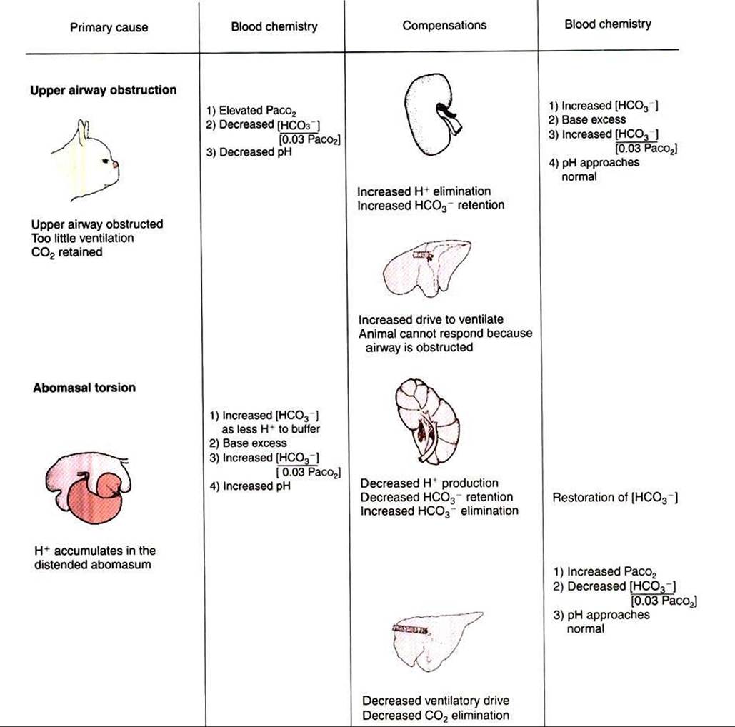
FIGURE 52-6 Diagrammatic representation of the acid-base changes initiated by upper airway obstruction, abomasal torsion, and neonatal foal diarrhea.
volume and cardiac output. To maintain blood pressure, vasoconstriction occurs in the extremities, which therefore have less blood flow and become cold. The loss of fluid from the interstitial space causes the dryness of the eyes and muzzle, the sunken appearance of the eyes, and inelastic skin. The increased hematocrit of 65 mL/dL (normal, 45) confirms the dehydration.
Feces contain HCO3", and its excessive loss causes a base deficit and a decrease in pH. In addition, poor tissue perfusion results in lactic acidosis. The acidosis results from loss of buffer base and accumulation of lactic acid; it is a metabolic acidosis. The acidosis stimulates ventilation, which reduces Paco2 in an attempt to correct pH. The foal has a metabolic acidosis that is partially corrected by the reduction in Paco2.
Treatment. This foal needs fluid replacement to increase plasma volume, raise cardiac output, and restore circulatory perfusion. I he fluid should contain electrolytes, to replace those lost in diarrhea, and a source of buffer, such as bicarbonate. A good choice would be lactated Ringer’s solution supplemented with additional bicarbonate. If the foals respiratory and acid-base homeostasis can be maintained
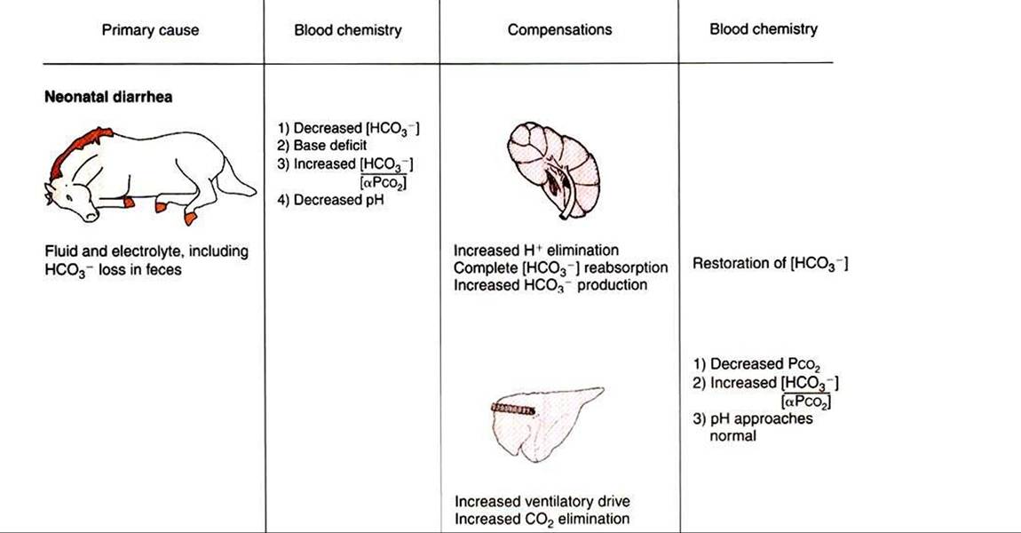
FIGURE 52-6 Continued.
until the diarrhea ceases, the foal has a moderate chance of recovery. A serious concern is that this 2-week-old foal could have an infectious cause of diarrhea. The foal could become septic from the infection, or the foal could have an initial septicemia, which is now manifested as diarrhea. In either situation, antibiotics are also warranted in most cases to treat the infection.