URINARY SYSTEM
The urinary organs function as with all mammals to remove waste products from the blood circulation, and any dysfunction can be life-threatening.
Kidneys
The kidneys are both retroperitoneal, lying in the sublumbar region on either side of the vertebral column, main aorta, and caudal vena cava (Fig.
12.16). They have a typical bean shape. They are typically mammalian with the usual cortical and medullary areas around the pelvis of the ureter. However, the autonomic nerve supply is more complicated than in other species (Whary & Andrews 1998).Serum chemistry in ferrets differs from dogs and cats in that the serum creatinine findings do not parallel elevations in the blood urea nitrogen (BUN) in renal failure (Hillyer 1997). Later estimations of glomerular function rate decided that using serum BUN and creatinine readings to determine renal insufficiency are questionable as the BUN can be influenced by non-renal factors. It is stated that the increase in serum concentrations of both substances do not actually appear until the kidney is 75% damaged (Esteves et al. 1994). It has been considered that creatinine elevation for ferrets is much lower than that of dogs and cats, as the normal mean value is lower (0.4-0.6 mg/dL) and the range is narrower (0.2-0.9 mg/dL). It may be that renal tubular secretion or enteric factors may be more prominent in affecting creatinine metabolism in ferrets than other animals (Rosenthal 1994). For laboratory ferrets with a food consumption of 140-190 g per 24 hrs, and a water intake of 75-100 ml per 24 hours, the urine volume is put at 26-28 ml per 24 hours with a urine pH of 6.5-7.5 (Fox 1998).
Enlargement of one or both kidneys has been recorded in the USA in ferrets over 3 years of age (Fox et al. 1998; Jenkins & Brown 1993). The condition can be associated, not with infection, but with renal cysts (Fig.
12.17). These can be single or multiple cysts and are considered to be hereditary, developmental or acquired (Dillberger 1995), usually with no clinical signs, or perhaps associated with renal failure (Rosenthal 1994). Acute and chronic renal failure, other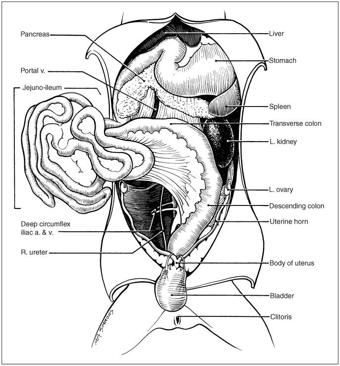
Figure 12.14 • Ferret abdominal viscera with intestines displaced. (Courtesy of Howard Evans.)
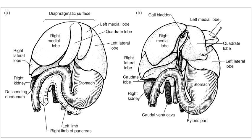
Figure 12.15 • (a) The liver showing organs in situ, (b) The liver reflected showing gall bladder and with pancreas removed. (Courtesy of Howard Evans.)
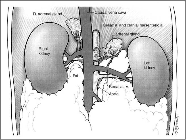
Figure 12.16 • Ferret adrenal glands and kidneys. (Courtesy of Lippincott Williams & Wilkins and Howard Evans.)
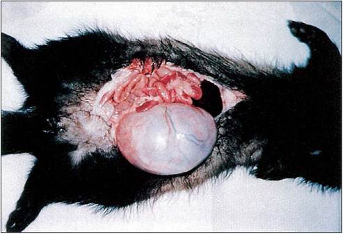
Figure 12.17 • Renal cyst in 3-month-old ferret. Slide courtesy of Dr. G. Rich.
Urine is used to mark territory by ferrets, as with other animals. An interesting point in bladder innervation is that sympathetic and parasympathetic nerves cause contraction of the whole bladder, while in other animals the sympathetic nerves produce relaxation (Whary & Andrews 1998).
CLINICAL NOTE
The bladder can be the site of cystic calculi, possibly due to commercial foods as it is not seen in ferrets fed fresh meat. Usually the condition is acute in males and chronic in females.
KEY NOTES
than cystic kidneys, can occur as with other mammals and treatment has been described (Lewington 2003c).
Ureters
The delicate ureters run from the renal pelvis and enter the base of the bladder from the left and right side. In the male ferret they are affected by inflammation of the prostate when urinary outflow is restricted.
Bladder
The bladder naturally varies in size depending on contents but when empty measures roughly 1 cm wide by 2 cm long. In the adult it has the capacity of around 10 ml of urine.
• The ferret has a large lung capacity relative to its size.
• Blood typing for transfusion between ferrets is not required as for other mammals.
• BUN and creatinine values differ from those in dogs and cats.