DIGESTIVE SYSTEM
Dentition
Adult ferrets have typical carnivore dentition with large curved canines and strong premolars and molars (Figs. 12.3b and 12.9).
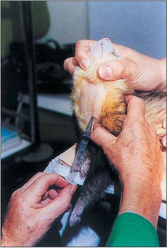
Figure 12.7 • Taking blood from the jugular vein.
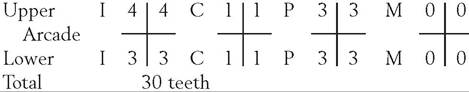
Photo courtesy of John Tingay.Deciduous dentition (Pass et al. 1993)
CLINICAL NOTE
The kitten's temporary teeth first erupt between the 3rd and 4th week so it is possible for needle-sharp canines to inflict damage on the jill's mammary glands, leading to mastitis.
Permanent dentition (Pass et al. 1993)
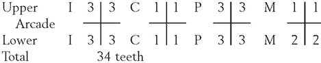
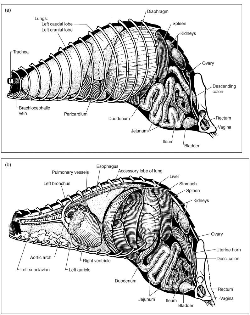
Figure 12.8 • (a) The thoracic and abdominal viscera by a superficial left lateral view. The dotted line shows the curve of the diaphragm. (b) The ferret thoracic and abdominal viscera on lateral view showing left lung removed. The dotted lines show the stomach passing to the pylorus dorsally and the duodenum. (Courtesy of Howard Evans.)
The ferret's permanent teeth appear from the 7th week of age, with the upper and lower canines plus the first lower molar appearing first. At about 53 days the upper molar is seen. This is followed by the second, third, and fourth upper premolars and the second and third lower premolars, which are all present by 67 days after birth. Finally, in the lower jaw, the fourth premolar and second molar are present a week later (Evans & An 1998).
The rostral view of the adult ferret skull (Fig. 12.9) illustrates the very narrow ventral space in the nasal conchae, through which only a 3.0 or 3.5 French red rubber catheter could be passed in an emergency.
The ferret or polecat jaw is powerful enough to kill small prey by crushing the skull, using the biting canines. A well- adjusted pet ferret is unlikely to bite to hurt unless alarmed or in pain. Young ferrets may nip in the process of “playfighting,” either with each other or with their owner, but the skin is not usually penetrated.
Abnormalities can occur in the dentition. Western Australian ferret kittens have shown supernumerary incisors. In one
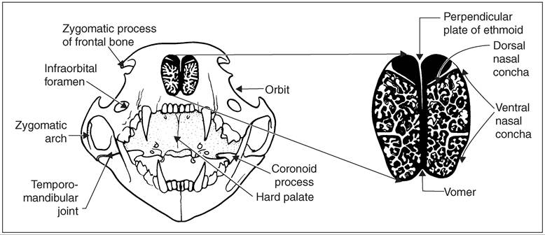
Figure 12.9 • Ferret skull (rostral view). (Courtesy
of Howard Evans.)
survey of 350 ferrets from various UK breeders, 26 ferrets had one or two supernumerary incisors (Andrews et al. 1979). There were three ferrets with broken canines. This was considered at the time an action by ferreters to stop them killing rabbits, but it is not necessary and today would be
regarded as a mutilation. Canine teeth can be fractured in fights or by accidents, and modern ferret dentistry can effect a repair (Johnson-Delaney & Nelson 1992). Basic dentistry is commonly carried out for scaling and extracting teeth under anesthesia with pet ferrets, as with dogs and cats.
243
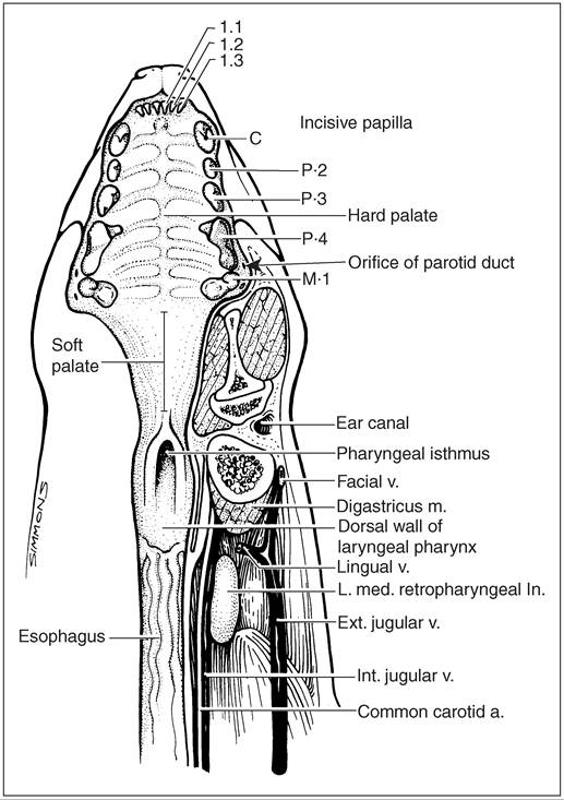
Figure 12.10 • Ferret pharynx and structures of interest. (Courtesy of Howard Evans.)
CLINICAL NOTE
In the upper dental arcade the first molar appears tucked behind the fourth premolar and is sometimes the site of a root abscess.
Muscles of mastication
The well-developed masseter muscle originates at the zygomatic arch and inserts on the masseteric fossa, condyloid crest, and mandibular angular process. The digastric muscle originates on the jugular process and tympanic bulla and passes to the ventral border of the caudal portion of the mandible and has the action of opening the jaw. The major adductor muscle of the lower jaw is the temporalis and this is well developed in the hob.
The deep pterygoid muscles, lateral and medial, assist the masseter and temporalis muscles in the crushing and chewing motion of closing the jaws.GENERAL INTEREST
The ferret has a powerful bite and can clamp its jaws tight on prey and will not let go. Large strong birds, which have been bitten on the foot, have been known to take weasels, stoats and even polecats aloft!
Tongue
The ferret tongue (Fig. 12.11) is long and freely movable and can be pulled forward to expose the tracheal entrance for endotracheal tubing, as in other mammals. The lingual frenulum can be the site of grass awn penetrations, especially in working ferrets in summer.
Salivary glands
The ferret has five pairs of salivary glands: parotid, mandibular, sublingual, molar, and zygomatic (Fig. 12.12). These glands can be damaged in fights between hob ferrets, typically in the mating season. The resulting formation of mucoceles will require surgical drainage. Miller and Pickett (1989) have described an operation on a zygomatic salivary gland mucocele.
Gastrointestinal tract
The esophagus can have a dilated transthoracic section, defined as a megaesophagus, which is sometimes also seen in puppies. This condition has occurred in ferrets but is now a rarity. The musculature of the esophagus is thin and weak and motility is reduced, leading to typical food bolus collection and regurgitation. Ferrets are able to vomit and have been used in experiments on the physiology of vomiting relating to humans.
The ferret has a short digestive tract like other carnivores, with a simple stomach lying on the curve of the liver in the cranial abdomen (Figs. 12.8a, b and 12.13). The stomach is capable of tremendous swelling and an adult hob ferret has been known to eat 80 g of meat at one time and then slowly digest it overnight.
Intestines
The small intestine is approximately 182-198 cm long and extends from the pylorus of the stomach to the junction with the colon (Evans & An 1998)(Figs.
12.13 and 12.14). The duodenum, the proximal loop of small intestine, is about 10 cm long. The ileum and jejunum have no apparent demarcation and pass to the large intestine, which is approximately 10 cm long. There is no ileocolonic valve in the ferret and no cecum or appendix. Because the cecum is missing, the ileocecal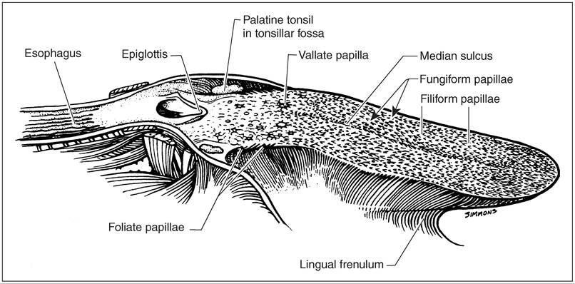
Figure 12.11 • The ferret tongue. (Courtesy of Howard Evans.)
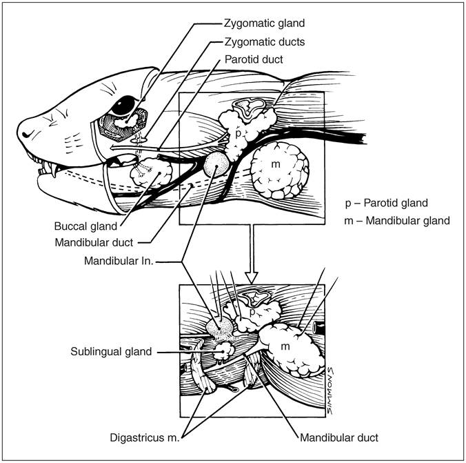
Figure 12.12 • Ferret salivary glands. (Courtesy of Howard Evans.)
junction is indistinct; however, the junction can be inferred by the pattern of the jejunal artery, which anastomoses with the ileocecal artery. The colon is divided into ascending, transverse, and descending portions and ends at the junction with the rectum at the pelvic inlet level (Evans & An 1998). The bowel is subject to obstructions: for example, chewed plastic toys. Foreign body operation procedure has been described (Bennett & Pye 2000).
The anus has an internal (smooth muscle) and external (voluntary muscle) sphincter system (Evans & An 1998). The external sphincter encloses the paired musk glands, which have openings on either side of the anal canal (Fig. 12.18). The musk glands are approximately 10 mm by 5 mm and their removal (anal sacculectomy) has been described (Bennett & Pye 2000).
Liver
(Fig. 12.15). The liver can be the site of primary neoplasia or subject to secondary invasions of malignant cells.
Pancreas and spleen
The pancreas is an elongate, lobulate, inverted ‘V'-shaped organ, usually light pink to bright red in color (Figs. 12.14 and 12.15). It can be the site of insulinoma cancers in ferrets. Delicate surgery for pancreatic beta-cell tumors has been described (Bennett & Pye 2000).
The spleen is a gray-brown organ lying in the left hypogastric area, running parallel to the greater curvature of the stomach.
It is crescent shaped and can become large quite normally in adults, though also very enlarged as a primary or secondary cancer.The diaphragm itself is divided into a typical muscular dome with central tendinous area and two crura. The liver fits into the mould of the ferret diaphragm and is relatively large compared to the average ferret body weight; an 800-1150 g animal could have a liver of 35-59 g (Evans & An 1998). The liver has right lateral, right, and left medial lobes, a quadrate central lobe hiding the gall bladder, and a left lateral lobe, all in the curvature of the diaphragm
KEY POINTS
• The ferret skeleton is lightweight but extremely flexible and strong.
• The thoracic cavity is large relative to body size.
• Ferrets have no ileocolonic valve, cecum, or appendix.
246
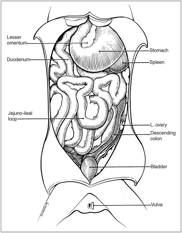
Figure 12.13 • Ferret internal viscera - undisturbed as for an abdominal operation. (Courtesy of Howard Evans.)