Uterus
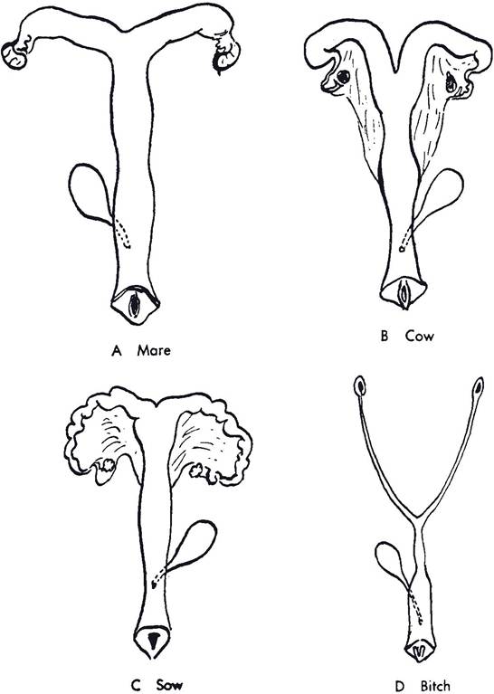
The uterus of the domestic mammal consists of a body, a cervix (neck), and two horns (Fig. 26-1). The relative proportions of each vary considerably with the species, as do the shape and arrangement of the horns (Figs.
26-4 and 26-5).Relative to the extent of the horns, the body of the uterus is largest in the mare, less extensive in the ruminant, and small in the sow. Externally, the body of the uterus of the cow appears larger than it is because the caudal parts of the horns are bound together by the intercornual ligament, which obscures their individual nature.
As with most other hollow internal organs, the uterine wall consists of a lining of mucous membrane, an intermediate smooth muscle layer, and an outer serous layer of peritoneum. The uterus is suspended bilaterally from the body wall by the mesometrium. The mesometrium, mesosalpinx, and mesovarium collectively constitute the broad ligament.
The mucosa lining the uterus, the endometrium, is a highly glandular tissue that varies in thickness and vascularity with hormonal changes in the ovary and with pregnancy (see Chapter 27). The epithelial covering of the mucous membrane is of the simple columnar type in the mare, but is stratified columnar epithelium in the sow and ruminants.
The uterine glands are simple branched tubular glands that exhibit considerable coiling. These glands are particularly active during estrus and pregnancy, during which they
Figure 26-4. Comparative shapes of animal uteri. The female dog (bitch) is provided for comparison.
produce a fluid colloquially known as uterine milk. These glands are scattered throughout the endometrium of the uterus, except in ruminants, in which the caruncles are nonglandular. Caruncles are mushroomlike projections from the inner surface of the uteri of ruminants; they provide a site of attachment for the fetal membranes (see Chapter 28).
The cervix of the uterus projects caudally into the vagina.
The cervix is a heavy, smooth muscle sphincter that is tightly closed except during estrus and parturition. During estrus the cervix relaxes slightly, permitting spermatozoa to enter the uterus. in ruminants, and to some extent in sows, the inner surface of the cervix is arranged in a series of circular ridges or rings, sometimes called annular folds (Fig. 26-6). The cervix of the mare is relatively smooth and projects prominently into the vagina, which surrounds the cervix as a deep vaginal fornix.The tunica muscularis is the muscular portion of the uterine wall, commonly called the myometrium. it consists of a thick inner circular layer of smooth muscle and a thinner outer longitudinal layer of smooth muscle, separated by a vascular layer. During pregnancy, the amount of muscle in the uterine wall increases dramatically, both in cell size (hypertrophy) and in cell numbers (hyperplasia).
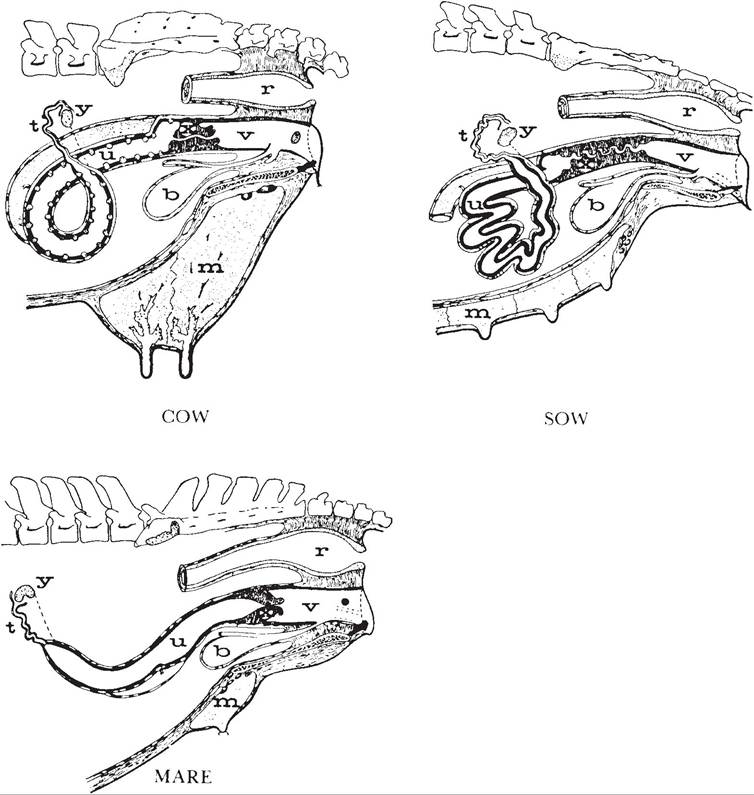
Figure 26-5. Comparative anatomy of the female reproductive tract. Lateral view. b, Urinary bladder; m, mammary gland; r, rectum; t, uterine tube; u, uterus; v, vagina; x, cervix; y, ovary. (Reprinted with permission of Wiley-Blackwell from Hafez, E.S.E. Reproduction in Farm Animals. 6th ed. Philadelphia: Lea & Febiger, 1993.)
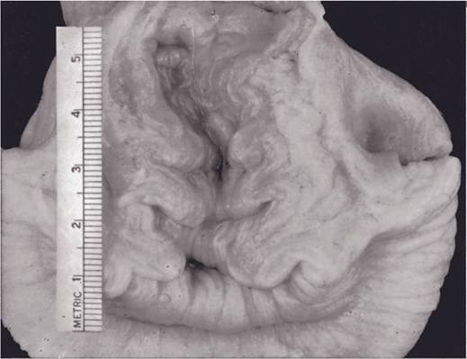
Figure 26-6. Cervix of a young cow, sectioned and opened to reveal the annular folds. (Reprinted with permission of Wiley-Blackwell from Hafez E.S.E. and Hafez B. Reproduction in Farm Animals. 7th ed. Philadelphia: Lippincott Williams & Wilkins, 2000.)