Vascularization
The relatively small ovarian artery, a direct branch of the aorta in cattle, supplies the ovary, the uterine tube, and the adjoining part of the horn of the uterus. The ovarian artery is distinguished by an extraordinarily convoluted course within the cranial part of the broad ligament and has extensive contact with the plexiform ovarian vein (Fig.
29.19). These features facilitate the transfer of prostaglandins from venous to arterial blood. The uterine artery arises from the internal iliac and enters the pelvic cavity within the broad ligament. It is ostensibly a branch of the umbilical but appropriates virtually the entire flow of its parent (Fig. 29.4). It is the largest of the arteries to the female tract, and before reaching the uterus, it divides into cranial and caudal parts, each the source of about half a dozen stem vessels that reach the mesometrial border of the uterus. Branches from these run over the uterine walls following courses that appear to coincide with the locations of the caruncles internally. The arrangement leaves the antimesometrial border of the uterus less well supplied and thus less prone to bleeding when incised. The vaginal artery, branching from the internal iliac near the ischial spine, runs over the dorsolateral surface of the vagina before swinging forward over the lateral wall, where it risks involvement, with possibly fatal outcome, in vaginal rupture, a relatively common calving catastrophe in heifers. Various branches pass to the caudal genital tract and to the bladder and urethra.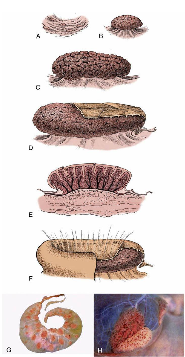
FIG. 29.17 Development of caruncles in the wall of the bovine uterus. (A) Caruncle in a nongravid
uterus. (B) Caruncle in a 2-week gravid uterus. (C) Caruncle in a 6-month gravid uterus. (D) Caruncle near term, covered in part by a cotyledon (fetal tissue). (E) Section of a placentome. (F) Placentome of a sheep. (G) Cotyledonary placenta (ruminant). (H) Partial separation of maternal and fetal parts of placentome (cow).
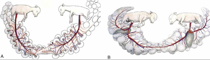
FIG. 29.18 (A) Twin bovine pregnancy showing separate circulations. (B) Twin bovine pregnancy showing conjoined circulations (freemartin development possible).
A very large and conspicuous venous plexus lies in the parametrial tissues of the broad ligament and over the ventral surface of the uterus and vagina, partly covered by the outer layers of muscle. It constitutes a blood pool that can drain in several directions (Fig. 29.19). The ovarian vein, the largest emissary vessel, runs in the cranial part of the broad ligament; the vaginal veins, including a surprisingly small vein that corresponds to the large uterine artery, play a secondary role. Both sympathetic and parasympathetic nerves supply the genital tract.