VASCULARIZATION AND INNERVATION
The reproductive organs are principally supplied by the ovarian, uterine, and vaginal arteries. The ovarian artery, a direct branch from the aorta, divides into uterine and ovarian branches.
The ovarian branch pursues a tortuous course within the mesovarium before dividing into several branches that spread over the surface of the ovary; this contrasts with the arrangement in other species, in which the vessels penetrate the ovary immediately on arrival. The other branch passes to the cranial part of the horn. The corresponding vein is disproportionately large and drains much of the uterus in addition to the ovary. Little transfer of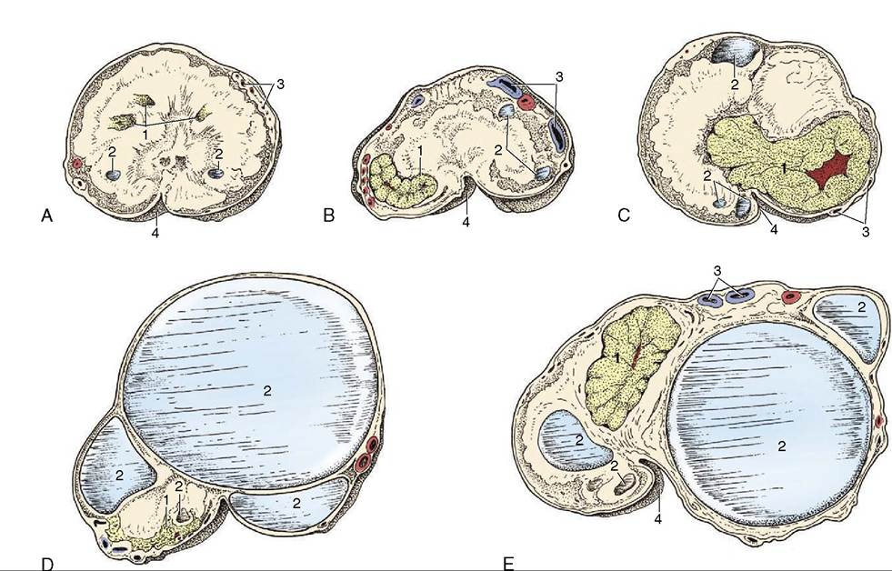
Figure 22-10 Sections of ovaries in various functional states. A, Ovary with corpora lutea and small follicles. B, Ovary with developing corpus luteum. C, Ovary with fully developed corpus luteum. D, Ovary with mature follicle. E, Ovary with follicles of various sizes and a rather large corpus luteum. The corpus luteum of the mare does not protrude from the ovary as in other species. 1, Corpora lutea; 2, follicles; 3, blood vessels; 4, ovulation fossa.
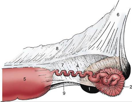
Figure 22-11 The right ovary, uterine tube, and uterine horn; lateral view. 1, Ovary; 2, infundibulum with fimbriae; 3, ampulla of uterine tube; 4, isthmus of uterine tube; 5, uterine horn; 6, mesovarium; 7, mesosalpinx; 8, mesometrium; 9, entrance to the ovarian bursa.
prostaglandins from venous to arterial blood occurs in the mare, which is a fact that may be correlated with the less intimate relationship of the ovarian artery and vein than exists in many other species.
The uterine artery, a branch of the external iliac, is the foremost supply to the uterus. It divides into several branches within the broad ligament, and these approach the mesometrial border of the horn and body separately. The antimesometrial aspect is reached only by small vessels, thus lending itself to relatively bloodless incision. Anastomoses with branches of the ovarian and vaginal arteries are present.
The vaginal artery takes origin from the internal pudendal in common with the middle rectal artery. It passes through the retroperitoneal tissue lateral to the vagina before bending forward to divide and supply the larger part of the vagina, the cervix, the caudal part of the body of the uterus, the bladder, and the urethra. The remaining part of the vagina and the vestibule are supplied from the vestibular branch of the internal pudendal artery.
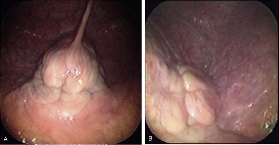
Figure 22-12 The changing appearance of the cervix. A, Dioestrus. B, Oestrus.
The veins draining the genital organs are satellite to the arteries. The innervation displays no noteworthy special features.