Adnexa
Chronic Blepharitis
Blepharitis, or inflammation of the eyelids, typically presents with clinical signs that vary based on etiology, chronicity, and degree of self-trauma but generally exhibits some degree of eyelid swelling, hyperemia, alopecia, crusting, and secondary keratitis and conjunctivitis (Pena and Leiva 2008) (Figure 4.1).
Some cases will present with ulcerated lesions either instead of or in addition to the classic swelling (Figure 4.2). Self-trauma from pruritis is often significant and may result in further damage to the skin, conjunctiva, and corneas that may confound both diagnosis and therapy. Chronic inflammation of the lids can result in eyelid distortion with cicatrix formation leading to either entropion or ectropion and secondary c orneal and conjunctival irritations (Pena and Leiva 2008; Bistner 1994). Damage to the meibomian glands or their ducts can impair the production and secretion of lipids from the eyelid glands resulting in a tear quality deficiency, rapid tear fluid evaporation, and clinical signs of exposure keratitis and dry eye (Samuleson 2013).There are innumerable causes of blepharitis, from infectious etiologies (parasites, bacteria, fungi, Leishmania) to allergies to immune-mediated disease, trauma, and neoplasia (Pena and Leiva 2008, Bistner 1994; Peiffer 1980; Lindley, Boosinger, and Cox 1990; Yamaki and θhono 2008). Diagnostics to rule out the more easily treated conditions would include skin scrapings and cytology to look for infectious agents and biopsy for histopathology and microbiologic culture and susceptibility. The cases that most commonly take on a protracted or chronic course are usually allergic or immune-mediated in nature. Animals with allergic blepharitis are
Chronic Disease Managementfor Small Animals, First Edition. Edited by W. Dunbar Gram, Rowan J. Milner and Remo Lobetti.
© 2018 John Wiley & Sons, Inc. Published 2018 by John Wiley & Sons, Inc.
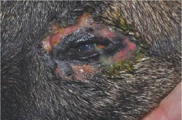
Figure 4.2 Immune-mediated blepharitis in a German Shepherd Dog. This case presented with ulcerated lesions rather than the nodular swellings seen in Figure 4.1 and had lesions at sites distant to the eyes.
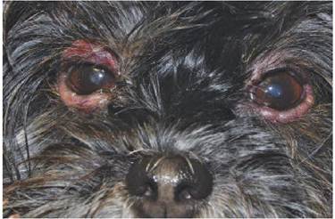
Figure 4.1 Chronic immune-mediated blepharitis in a mixed-breed dog. This case was steroid responsive.
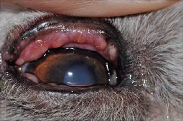
Figure 4.3 Meibomianitis in a Shih Tzu. Note the accumulations of glandular secretions in the subconjunctival space.
usually pruritic and also exhibit signs distant to the eyes, especially the ears and feet (Pena and Leiva 2008). Treatment of any secondary infections should be initiated along with allergy testing and elimination therapy or desensitization therapy to address the underlying cause. Supportive care with topical anti-inflammatories for the eyelids and the associated conjunctivitis is usually helpful and may be required intermittently for flare- ups. Immune-mediated blepharitis can take several forms and may be accompanied by ocular or multi-organ disease (Pena and Leiva 2008; Bistner 1994; Peiffer 1980; Lindley, Boosinger, and Cox 1990; Yamaki and Ohono 2008). The pemphigus complex diseases, discoid and systemic lupus erythematosus, and uveodermatologic syndrome are examples of autoimmune conditions that often affect the eyelids as well as distant sites, often with depigmentation, ulceration, and crusting (Pena and Leiva 2008; Lindley, Boosinger, and Cox 1990; Yamaki and Ohono 2008). Biopsy is required to confirm diagnosis of these conditions and chronic (often life-long) systemic therapy is necessary. Uveodermatologic syndrome, sometimes referred to as Vogt-Koyanagi-Harada syndrome, is a condition in which tissues heavily-laden with pigment (melanin) are the target of disease and the uveitis that develops is usually severe and can lead to secondary glaucoma and vision loss (Lindley, Boosinger, and Cox 1990; Yamaki and Ohono 2008).
These immune-mediated diseases require systemic therapy with immunosuppressive or immune-modulating therapy.Meibomianitis is an inflammation of the meibomian glands located within the eyelids at the margins (Samuleson 2013). Inflammation of these glands causes eyelid swelling and often an observable enlargement of the meibomian glands and chalazions seen when the eyelids are everted to reveal the palpebral conjunctiva (Figure 4.3). This condition is often secondary to a bacterial infection or hypersensitivity. However, it can become chronic and will lead to reduction of tear film lipid and ocular surface inflammation and damage. Topical and systemic antibiotics, selected based upon culture of the exudate from the glands, are indicated as well as systemic corticosteroids if an allergic or immune-mediated etiology is suspected. Warm compresses may provide some relief to the patient.
Eyelid Neoplasia
Neoplasia of the eyelids is very common in dogs, less so in cats. Dog eyelids exhibit a variety of neoplasms of the eyelids that are usually benign and typically responsive to conservative surgical procedures. Many small eyelid tumors are slow growing and do not initiate any clinical signs. Others, however, may cause a considerable amount of ocular surface irritation that will become chronic if not addressed. Meibomian adenomas, epitheliomas, and papillomas are the most common types of neoplasia, however, melanomas, mast cells tumors, basal cell tumors, and fibroma/ fibrosarcomas are noted occasionally (Aquino 2007; Roberts, Severin, and Lavach 1986).
Eyelid neoplasia in cats, however, tends to be aggressive and often malignant, requiring intervention before a chronic condition can be established. Squamous cell carcinoma is by far the most common type of eyelid neoplasia in the cats, most often seen in older felines with light-colored hair coats and minimally pigmented skin (Aquino 2007; Roberts, Severin, and Lavach 1986). The lesions tend to be erosive or ulcerated wounds rather than proliferative masses. Early intervention is recommended to minimize local extension. Other tumors that have been reported in the eyelids of cats include mast cell tumors, papillomas, adenoma/adenocarcinomas, basal cell tumors, fibrosarcomas, and peripheral nerve sheath tumors (Aquino 2007; Roberts, Severin, and Lavach 1986).