Anatomy
The pancreas in dogs and cats is a long narrow structure that can be divided into a right and a left limb. In between these two limbs is the head of the pancreas, which, in contrast to humans, is not an easily discernible structure in dogs (Figure 8.1) and cats.
The right limb of the pancreas lies directly next to the duodenum, while the left limb is situated next to the spleen. The pancreas is made up of lobules of pancreatic tissue, which in turn are made up of acinar cells (Figure 8.2). In between the lobules there are the islets of Langerhans (Figure 8.2), which are collections of neuroendocrine cells. These neuroendocrine cells make up the endocrine portion of the pancreas and synthesize and secrete a variety of regulatory polypeptides, most importantly insulin and glucagon. The acinar cells produce digestive enzymes and zymogens, which are released into the duct system that leads to the duodenum.Dogs usually have two pancreatic ducts. The main pancreatic duct, ductus pancreaticus, empties into the duodenum on the major duodenal papilla together with the common bile duct. The lumen of the pancreatic duct is separated from the duodenum by the sphincter of Oddi, which is a muscular sphincter that is essential in preventing duodenal contents from entering the pancreatic duct. Dogs and approximately 20% of cats have a second pancreatic duct, the accessory pancreatic duct (ductus pancreaticus accessorius) that empties on the minor duodenal papilla, approximately 1-3 cm distal to the major duodenal papilla into the duodenum.1
Table 8.1: Secretory products of the exocrine pancreas
This table shows a list of secretory products of the exocrine pancreas and their main functions. There are three types of secretory products: zymogens of pancreatic digestive enzymes, pancreatic enzymes, and other molecules that are neither.
| Enzymes secreted as zymogens | Enzymes secreted in an active form | Other secretory products |
| trypsinogen | lipase | water |
| chymotrypsinogen | amylase | bicarbonate |
| proelastase | carboxylesterase | procolipase |
| prophospholipase | desoxyribonuclease | intrinsic factor |
| kallikreinogen | ribonuclease | antimicrobial factors |
| procarboxypeptidase | pancreatic secretory trypsin inhibitor (PSTI) trophic factors for the intestinal tract |
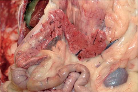
Figure 8.1:
Normal canine pancreas. This figure shows a normal canine pancreas collected during necropsy. Note the right and left limbs on either side of the head of the pancreas. In dogs and cats, the head of the pancreas is not as clearly demarcated as is the case in humans, but it can be identified by the pancreatic artery, vein, and duct that enter the pancreas in this area. (Image courtesy of Dr. Shelley Newman, University of Tennessee, USA.)
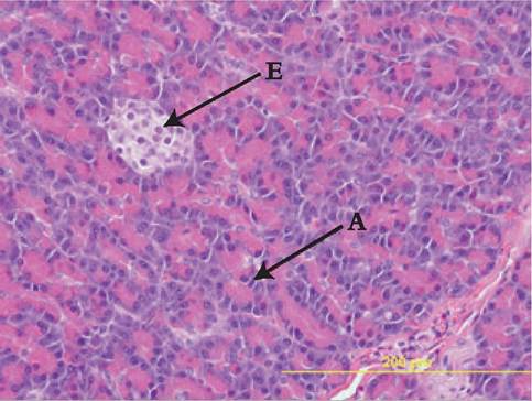
Figure 8.2:
Histological view of the pancreas. This figure shows a histological view of a normal dog pancreas. The majority of the cells are exocrine pancreatic cells (A) arranged in acini that subsequently form the pancreatic lobules. The islets of Langerhans are composed of clusters of endocrine cells (E), which have smaller nuclei and a more vacuolated cytoplasm. (H&E stain, 40?; image courtesy of Dr. Shelley Newman, University of Tennessee, USA.)
8.2