Anemia
Guy D. Lester
Anemia is a common finding in neonatal foals. The normal ranges of hematologic parameters must be considered when determining the significance of any anemia. The packed cell volume (PCV) at birth is 40% to 52% but decreases to 32% to 46% after colostrum and milk intake by 24 hours of age.
PCV, red blood cell count (RBC), and the hemoglobin (Hb) concentration are similar to adult values at 24 hours of age, before decreasing over the first 2 weeks of postnatal life to values that are in the lower level of the normal adult ranges. These relatively low levels are sustained for several months. The mean corpuscular volume (MCV) decreases over the first 4 months of life, leading to microcytosis and anisocytosis. The MCV increases after weaning.281,282 Foals typically have reduced serum ferritin concentrations and increased total iron binding capacity when compared with adults. Serum ferritin increases significantly on day 1 and is attributed to ingestion of colostral iron, before reducing. Data support relative iron deficiency as a cause of common foal anemia, but supplementation with iron does not improve red cell indices. Supplementation of normal foals with a specific iron preparation, ferrous fumarate, resulted in acute hepatic failure and death. There are reports of absolute iron deficiency anemia in foals, including stabled Dutch Warmblood foals fed cut grass. Findings include a low PCV, low serum iron and ferritin concentration, and normal to increased total iron binding capacity. Improvement is seen in those foals with oral iron supplementation.Clinical signs of anemia are related to the magnitude and timing of the underlying disease process. Rapidly developing hemolytic anemias, such as NI, produce signs of weakness, lethargy and somnolence, mucous membrane pallor and icterus, tachycardia, and possible mild fever.
Intravascular hemolysis will typically cause brown-red discoloration of urine. There is accentuation of cardiac murmur. Foals with hemoperitoneum or internal umbilical remnant hemorrhage are often colicky. Hemorrhage at the apex of the bladder can produce stranguria. Bleeding into the respiratory cavity can produce clinical signs of respiratory distress, and blood loss into the gastrointestinal tract will cause melena. Lameness will be seen in foals with spontaneous hemarthrosis.Causes
sulfonamides, pyrimethamine, folic acid, and vitamin E. There is a syndrome of anemia and B-cell lymphopenia in Fell pony foals. Affected foals demonstrate initial bone marrow erythroid hyperplasia with concurrent erythroid and myeloid dysplasia, which progresses to severe erythroid hypoplasia by 5 weeks of age.
Determination of the nature of the anemia may allow specific treatment. The treatment of NI is discussed in Chapter 53. Drug-induced or autoimmune anemias may be treated with corticosteroids (0.05 to 0.1 mg/kg dexamethasone twice a day intramuscularly or intravenously). Blood transfusion following crossmatch may be indicated when anemia develops rapidly or PCV drops below 12%. Associated conditions, such as metabolic acidosis and hypoglycemia, should be corrected. Anemia of chronic disease requires correction of the primary disease condition.
Maturity
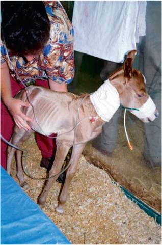
FIG. 17.11 A premature foal demonstrating a low birth weight and small body size, a short and shiny hair coat, and a prominent rounded head.
flexor laxity with elevation of the toe, but some will have contracture of the lower limb. Muscle development is usually poor. Most will demonstrate generalized weakness and hypotonia and will have difficulty in standing. Severely premature foals may have lids naturally sutured closed and little hair covering of their body. Many will have difficulty in maintaining body temperature, blood pressure, and blood glucose.
Dysmature foals commonly experienced some degree of intrauterine growth retardation (IUGR). This is usually reflected by the birth of a foal that is small for its gestational age. The average relative weight of the term foal to its dam is around 10%. Postmature foals usually have an acceptable birth weight with a large frame but poor muscle development. This gives the foal a lanky appearance. In contrast to premature animals, fetlock contracture is common, although laxity can be present. Consistent with their prolonged gestation, post-term or postmature foals will often have erupted incisors and a long hair coat. In term foals the central incisors typically erupt during the first 5 to 7 days of postnatal life.
Causes
The pregnant uterus is highly responsive to contractile agents such as oxytocin and prostaglandins throughout gestation. Consequently, one of the most important causes of premature birth and perinatal morbidity and mortality is the induction of labor with exogenous oxytocin or prostaglandins. The adverse consequences of premature induction of parturition were identified in a study where parturition was induced either before 300 days' gestation or between 300 and 320 days' gestation. The overall survival rate was only 5%, with the youngest surviving animal delivered after 318 days' gestation. Other surviving foals were all delivered after 320 days' gestation. The decision to prematurely terminate a pregnancy may be done deliberately in the “normal” mare or may be forced due to significant maternal disease. The latter frequently involves delivery of a compromised and often premature foal by cesarean section. Chemical induction of parturition has occurred when late-pregnancy intestinal problems are misinterpreted as ineffective labor. Premature birth can occur as a sequel to placental problems, including placental infection, edema, and/or detachment (premature placental separation). Placental insufficiency due to twinning is another cause of intrauterine growth retardation.
The consumption by pregnant mares of tall fescue pasture infected with the Neotyphodium Coenophialum leads to range of abnormal signs including prolongation of gestation, perinatal mortality, and agalactia. The large skeletal frame of the postmature foal predisposes mares to dystocia. The delay in parturition may be due to toxin-induced interference with fetal corticotrophin-releasing horses (CRHs) and delay in maturation of the HPA axis. Foals born to mares grazing endophyte-infected fescue pasture have normal thyroxine and reverse T3 but reduced tri-iodothyronine levels compared with control foals. This is also consistent with failure of cortisol- induced maturation of thyroid function. A syndrome of congenital hypothyroidism has been reported in foals in Western Canada. Signs include prolonged gestation, dysmaturity, and a range of musculoskeletal abnormalities, including flexural deformities, delayed ossification, and mandibular prognathism. The specific cause has not been determined, although consumption of diets that contain nitrate or are deficient in iodine is suspected.
Maturation of the Fetal Hypothalamic-Pituitary-Adrenal (HPA) Axis significant fetal cortisol appears to occur during the final 48 to 72 hours of pregnancy in mares. A number of important maturational events appear to be tightly associated with the prepartum increase in ACTH and cortisol. These include changes in red and white blood cell parameters, most notably 313314 a large increase in the neutrophil to lymphocyte ratio.313, Hepatic and renal glucose-6-phosphatase, a key enzyme of gluconeogenesis, also increases sharply around the time of birth coinciding with increases in hepatic and skeletal muscle glycogen stores.
The prepartum rise in plasma cortisol likely induces deiodination of the outer ring of T4 to produce the biologically active tri-iodothyronine (T3). Adequate levels of T3 are required for a number of biological functions including postnatal thermogenesis.
Normal term foals have high levels of thyroid hormones, including T3 at the time of birth.317,318 These levels decline over the initial weeks or months of postnatal life. A relationship between circulating T3 levels and cortisol was reported in premature, dysmature, and mature foals, and the increase in T3 appears to be dependent on maturation of the HPA axis.302,319 Both cortisol and T3 are critical for lung maturation, particularly the normal postpartum reabsorption of lung liquid.Accelerated Maturation of the Fetal HPA Axis
Several factors can induce premature maturation of the fetal HPA axis. Hypoxemia is a potent stimulator of the axis in sheep with rises in fetal ACTH and cortisol. The HPA axis can also be manipulated using exogenous glucocorticoids; betamethasone is commonly administered to women in danger of preterm birth in order to hasten HPA maturation and therefore improve the chances of postnatal survival. Poor nutrition before and after conception in sheep produces a shortened gestational period and hastened maturation of the fetal HPA axis. Placental and/ or fetal infection also can accelerate maturation of the HPA axis. The cytokines induced by infection increase prostaglandin synthesis and decrease metabolism. Prostaglandins exert a range of actions in addition to promoting cortisol production. These include important actions on the cardiovascular, renal, and respiratory systems. The healthy newborn foal has higher concentrations of 15-ketodihydro-PGF2α (PGM) in the days after birth than values in adult animals.
The stimuli associated with precocious HPA axis maturation in foals are not well described. The exception is infection of fetal membranes where foals are often delivered preterm with laboratory findings consistent with axis maturation. Spatial and nutritional deprivation resulting from a Thoroughbred foal placed in a pony uterus using embryo transfer also leads to premature maturation of the fetal adrenal.308,322,323 Many late-term maternal diseases do not appear to have a significant effect on foal maturity.
Hypoxemia associated with anesthesia and colic surgery in pregnant mares during the final 60 days of pregnancy results in a high rate of preterm delivery of compromised foals that did not survive. It is likely that the insult in these cases was so severe that the interval between surgery and delivery was inadequate for maturation of the axis to occur. Another important consideration in determining outcomes would be the effect of hypoxia and/or ischemia on other fetal organ systems.Treatment of the at-Risk Late Pregnant Mare
The administration of corticosteroids to the pregnant mare appears to have little effect on maturation of the fetal HPA axis, at least when doses considered safe are used. Direct injection of ACTH1-24 to the fetus results in increased fetal cortisol, but the effect is dependent on gestational age with maximal responses occurring at around day 313 with no measurable benefit when administered before day 295.313 Direct administration of CRH, ACTH, or betamethasone to the fetus using US-guided IM injection results in increased maternal progestagen levels consistent with maturation of the fetal adrenal gland.325,326 The procedure itself can lead to abortion in a small number of mares. Exogenous ACTH1-24 administered to late- pregnant pony mares had an impact on both gestational length and fetal maturation.327 Depot ACTH1-24 given to mares at 300, 301, and 302 days of gestation produced a shortened gestational length and lower birth weight but evidence of HPA axis maturation. A confounding effect in this study was the time of conception, with the most significant findings observed in mares bred later in the breeding season.
It is preferable to maintain the fetus in utero to ensure not only adequate HPA axis maturation but also effective ossification and maturation of other body systems. Consequently, the primary focus of therapy for a mare with placental infection is to eliminate the pathogenic organisms, reduce inflammation, and maintain the pregnancy. The specific management of placentitis is not the focus of this chapter, but treatment may involve broad-spectrum antibiotics, nonsteroidal antiinflammatory drugs, pentoxifylline, β2-adrenoreceptor agonists, and altrenogest. The efficacy of altrenogest use in mares with 328
placentitis has been questioned.328
The termination of post-term pregnancies is a difficult decision for practitioners, particularly with the angst that is common in many owners. Given the wide variation in gestational range, it is almost always in the best interests of the foal to let the pregnancy continue. If facilities are available, then rectal and transabdominal US assessment of the fetus and chorioallantois should be made, looking carefully for thickening or detachment of the fetal membranes. Ideally, any induction of parturition should be based on appropriate changes in physical characteristics of the mare and in milk electrolytes. Mares grazing endophyte-infected tall fescue can be medicated with dopamine receptor antagonists, such as domperidone.304
Laboratory Assessment
The laboratory data will indirectly reflect the degree of HPA axis maturation. Premature or dysmature foals that fail to survive often have minimal cortisol secretion in the face of adequate endogenous ACTH. Furthermore, the change in plasma cortisol in response to exogenous ACTH (Cosyntropin, 0.125 mg IM) is inconsistent and usually inadequate. The foal with incomplete adrenal maturation will have a low total white cell and neutrophil counts, as well as a neutrophil-to-lymphocyte ratio that is characteristically less than 1 : 1.314 It is important to determine if sepsis is present, as neutropenia is also a common feature of this condition. Evidence of shifting toward immature cell types and neutrophil toxicity should indicate primary sepsis or prematurity/dysmaturity complicated by sepsis. Premature foals that fail to improve their total white blood cell and neutrophil counts over the initial 24 to 48 hours of treatment have an even poorer prognosis for survival. Changes in red cell indices have also been reported in nonstressed preterm foals.314 Most notably is an elevated MCV in preterm foals. An elevated plasma fibrinogen concentration is considered to be a good prognostic factor in premature foals as it often reflects prepartum exposure to bacterial infection. Induced or spontaneously delivered term foals have a significantly higher plasma glucose concentration than premature foals.329 Plasma creatinine levels are often elevated in newly born preterm or dysmature foals due to placental dysfunction. This increase is independent of foal renal function. Measurement of low cortisol levels coupled with increased progestagens would provide further evidence that effective maturation of HPA axis has not occurred in the foal before birth.216 Thyroxine (T4) is lower in premature foals than in normal foals and term, but sick, hospitalized foals.317 This is consistent with failure of cortisol- induced maturation of thyroid function (see earlier). Premature foals respond to thyrotropin-releasing hormone (TRH; 0.5 mg IV), but the increase in T4 is less than that observed in both healthy and sick term foals.317
Establishing a Prognosis
The prognosis for survival of a prematurely delivered foal is dependent on a range of factors including the gestational age, reasons for delivery, complications associated with delivery, available resources (facilities and expertise), and financial limitations of the owner. Survival of very premature foals (280 to 300 days) would typically require a history of chronic in utero stress with resultant precocious maturation of the HPA axis and critical organ systems. The majority of these foals would still require a lengthy and costly period of hospitalization and experience a range of complications, some of which may be life threatening. Foals delivered prematurely as a consequence to chemical induction of parturition without evidence of chronic in utero stress or through cesarean section typically have a high mortality rate even when delivered close to calculated due dates. Foals delivered under these circumstances before 300 days will almost certainly die irrespective of available resources.
A complete blood count and fibrinogen estimation are key factors in determining short-term prognosis. A normal or elevated neutrophil count, neutrophil-to-lymphocyte ratio (N:L), or total white blood cell count are positive indicators of survival, as they typically reflect maturation of the HPA axis. In a survey of 135 neonates admitted to the University of Florida with a gestational age of fewer than or equal to 320 days' short-term survival was in part predicted by total white blood cell count, neutrophil count, lymphocyte count, and the N:L ratio at presentation.330 The N:L ratio of surviving premature foals (12.5 : 1) was well above that reported for both normal term foals (2.5 : 1) and that of nonsurviving premature foals. Many of the surviving animals were exposed to confirmed or suspected placental infection. In utero stress, with hastened maturation of the HPA axis, is a good prognostic factor for survival in foals that are delivered preterm. In many foals, the greater the neutrophil count, the better the outlook, at least in terms of short-term survival. A high plasma fibrinogen concentration is also considered to be a positive factor. Reevaluation of the white cell indices on day 2 for appropriate increases also support a favorable prognosis.
A history of placental infection appears to be a positive factor when predicting survival in preterm foals. One obvious downside is that many of these foals are born with aspiration pneumonia (due to in utero aspiration of contaminated amniotic fluid) and/or systemic sepsis. This, coupled with the fact that many foals have an impaired immune system, warrants the use of broad-spectrum antimicrobial therapy.
Consideration should also be given to the long-term outcomes of preterm foals. These animals are at risk of significant and permanent musculoskeletal problems due to bone and ligamentous immaturity. Foals that survive the neonatal period are smaller than their peers, and this difference will often remain noticeable as weanlings and as yearlings. The differences may be less obvious at 2 years and older. Other common complications, such as pneumonia, will further reduce the growth rate in the first 6 months of life. There is nothing to indicate that premature fillies will experience fertility problems as adults.
Clinical Progression
The clinical progression will usually reflect the degree of endocrinologic maturity, additional perinatal stresses, and the extent of physical maturity. Typically foals born prematurely,
■ TABLE 17.4
Normal Blood Gas Values for Foalsa
| Age Group | Arterial | Venous | |||||||
| O2 (mm Hg) | CO2 (mm Hg) | pH | Base Excess | hco-3 (mEq/L) | O2 (mm Hg) | CO2 (mm Hg) | pH | IICO 3 (mEq/L) | |
| Immediate | 40-50 | 52-60 | 7.2-7.3 | +2 | 24-26 | — | — | — | — |
| post-foalingb 2 hoursb | 68 ± 10 | 49 ± 2 | 7.37 ± 0.01 | +4 | 26 ± 2 | 42 ± 2 | 56 ± 2 | 7.33 ± 0.01 | 28 ± 2 |
| 4-12 hoursc | 75 ± 5 | 47 ± 2 | 7.39 ± 0.01 | +6 | 28 ± 2 | 42 ± 2 | 52 ± 2 | 7.38 ± 0.01 | 30 ± 2 |
| 24 hoursc | 81 ± 6 | 48 ± 2 | 7.4 ± 0.01 | +6 | 28 ± 2 | 42 ± 2 | 52 ± 2 | 7.38 ± 0.01 | 30 ± 3 |
| 1-3 daysc | 90 ± 6 | 148 ± 2 | 7.4 ± 0.01 | +6 | 28 ± 2 | 43 ± 2 | 52 ± 2 | 7.38 ± 0.01 | 29 ± 2 |
| 4-14 daysc | 86 ± 5 | 45 ± 2 | 7.41 ± 0.01 | +6 | 28 ± 1 | 38 ± 2 | 53 ± 2 | 7.38 ± 0.01 | 31 ± 2 |
| Prematureb birth | 39 ± 5 | 55 ± 4 | 7.27 | -3 | 24 ± 1 | — | — | — | — |
| (320-330 days’ | 52 ± 4 | 48 ± 3 | 7.33 | -1.3 | 25 ± 1 | — | — | — | — |
| gestation)— 1 hour | |||||||||
aLateral recumbency.
bData from Rose RJ, Rossdale PD, Leadon DP: Blood gas and acid-base status in spontaneously delivered, term-induced and induced premature foals. J Reprod Fertility 32:521, 1982.
cData from Stewart JH, Rose RJ, Barko AM: Response to oxygen administration in foals: effect of age, duration and method of administration on arterial blood gas values. Equine Vet J 16:329, 1984.
but chronically exposed to an appropriate chronic in utero stress, such as placental infection, will appear weak and depressed in the immediate postpartum period. Some will require resuscitation. After a longer than normal period of postural adaptation, they will usually manage to stand but will often require assistance. Suckle reflex and appetite may be reduced or absent, and many will need to be fed initially via nasogastric tube. They will frequently have trouble maintaining their body temperature and blood glucose levels. After the initial 24-hour period, many of these foals demonstrate improvement both in physical strength and mentation. Their appetite for milk will often exceed that of a healthy term foal. Foals with inadequate maturation of the HPA axis will frequently require immediate resuscitation. They may mimic the clinical progression of in utero stressed premature foals up until 12 to 18 hours of age. This initial period after delivery can be deceptive, with many foals showing degrees of improvement, which often promote owner optimism. The rise of hormones accompanying delivery may lead to improvement in alertness and strength. However, after this period a range of progressive abnormalities develop. These include systemic weakness, depression, seizures, respiratory failure, and intolerance to feeding. Cardiovascular collapse may ensue, the first sign of which is a reduction in the intensity of peripheral pulses, followed by a reduction in urine flow, development of subcutaneous edema, and deteriorating neurologic function. Poor tissue perfusion leads to lactate accumulation and a mixed metabolic and respiratory acidosis. Death will certainly occur without aggressive support, and even with high-level intensive care, mortality rates are high.
Treatment of the Premature or Dysmature Foal
Successful outcomes are dependent not only on careful management of identified problems but also in predicting the problems that may arise in the hours, days, or weeks to come. Most premature and dysmature foals will experience some degree of pulmonary insufficiency. Factors that predispose these foals to respiratory problems, including structural and functional immaturity, a naive and potentially immature immune system, altered pulmonary vascular reactivity, a highly compliant rib cage, and a propensity for prolonged or persistent recumbency. Final maturation of the respiratory system appears to be highly dependent on a functional HPA system. Arterial blood gas analysis is an important tool in the assessment of respiratory function, and the lower arterial oxygen concentration in term newborn foals is further decreased in dysmature or premature foals (Table 17.4). Extrapulmonary shunts account for more than 30% of the cardiac output, in contrast to less than 10% in normal full-term foals.331 Ventilation/perfusion mismatching also occurs due to a poorly reactive pulmonary vasculature and dependent atelectasis. Deficiency of lung surfactant is not likely to play a primary role in the respiratory dysfunction in most premature or dysmature foals, as it is usually fully developed in most foals by 300 days but could be delayed until after 340 days in some foals.332 The most severe form of respiratory failure is neonatal respiratory distress syndrome (RDS), a disease characterized by progressive respiratory failure, severe hypoxemia and hypercapnia, and death. A diffuse, severe alveolar pattern is a classical radiographic finding. Intervention would ideally involve mechanical ventilation, bovine or synthetic surfactant, and glucocorticoids; however, outcomes are extremely poor irrespective of the level of care. Fortunately, RDS is relatively uncommon; most premature foals will demonstrate a less severe manifestation of lung dysfunction, characterized by reduced ventilation capacity, tachypnea, hypoxemia, and varying levels of hypercapnia. These foals are susceptible to dependent lung atelectasis from recumbency. Most foals will benefit from supplemental intranasal oxygen with initial flow rates of 5 L/min recommended. Adjustment in flow rate is dictated by positive changes in arterial blood gas analyses or improvement in ventilation rate and depth. It is important to avoid prolonged periods of lateral recumbency in order to minimize the impact of atelectasis. If the foal is unable to stand, then placement in sternal recumbency is recommended. This is made easier by use of a specially constructed V-pad.
Failure of the cardiovascular system is common in foals with partial or incomplete maturation of the HPA axis. Management is challenging, in part due to inconsistent responses to standard inotrope and vasoreactive therapy (see earlier). Successful treatment is reliant on early detection of reduced perfusion. This is reflected clinically by cool extremities, the presence of limb and ventral edema, and darkening of the mucous membranes with prolongation of the capillary refill time. As failure ensues, peripheral pulses will become difficult to palpate, blood pH will fall, and there will be increases in plasma lactate and anion gap. Indirect (or direct) measurement of mean blood pressure along with determination of blood lactate will help guide therapy. An initial approach to the treatment of failing perfusion may involve intravenous plasma followed, if necessary, by dopamine and/or dobutamine infusion. Dopamine is widely available and relatively inexpensive, with effects that are dose dependent; (1) low doses (0.5 to 5 μg/kg/min) provide agonism of dopaminergic receptors and possible renotubular effects along with vasodilation of coronary and intestinal vasculature; (2) moderate doses (4 to 10 μg/kg/min) stimulate β1-adrenergic receptors and result in chronotropy; and (3) high doses (≥10 μg/kg/min) cause agonism of α1-adrenergic receptors, leading to widespread vasoconstriction. High-dose dopamine infusion is generally not recommended in human preterm infants due to the potential for right-to-left shunting.333 Dobutamine (3 to 20 μg/kg/min) acts principally on β1 adrenoreceptors, producing an improvement in myocardial contractility without vasoconstriction. Other potential pressor agents used in foal intensive care facilities include epinephrine, norepinephrine, and vasopressin. Milrinone infusion is used in human preterm infants and has been evaluated in anesthetized adult horses, but it has not been evaluated in premature foals.333,334 The volume and type of fluids given should be carefully monitored, as fluid overload and hypernatremia are common. Urine output should be appropriate for the volume of fluids administered and anuria or oliguria treated aggressively. This may include low-dose dopamine or fenoldopam infusion, furosemide boluses or infusion, or mannitol infusion. Establishment of urine flow is critical in terms of survival.
Signs of gastrointestinal tract dysfunction are rarely evident on initial assessment of premature or dysmature foals; however, most will not tolerate aggressive force-feeding. These foals commonly develop intestinal stasis with reduced fecal passage, gas accumulation, and gastric distension. The combination of prolonged asphyxia and prematurity is also a risk factor for the development of necrotizing enterocolitis. Feeding should be restricted to small volumes (e.g., 10 to 20 mL hourly) until the foal appears to be systemically stable. Concurrent parenteral nutrition is indicated in order to prevent loss of body weight. Foals should be monitored closely for signs of gastrointestinal dysfunction irrespective of feeding volume or frequency. This includes assessment of fecal passage, changes in abdomen size (assessed using a measuring tape), testing for gastric reflux if a nasogastric tube is in place, and frequent assessment with transabdominal US.
Premature and dysmature foals are susceptible to hypothermia. Thermogenic mechanisms develop late in gestation and are related to circulating T3 levels. As discussed earlier, thyroid hormone generation is closely tied to maturation of the HPA axis. Consequently, problems with thermogenesis are exacerbated in preterm foals with incomplete adrenal function. Body temperature needs careful management as rapid warming may result in peripheral vasodilatation and possible cardiovascular collapse. Initially, the foal should be covered by blankets and removed from any drafts. Intravenous and oral fluids should be warmed before use. Once the foal begins to demonstrate vigor, heat lamps and circulating warm-water blankets can be used. The premature or dysmature foal often has inadequate gluconeogenic enzyme activity and limited glycogen stores at the time of birth. Consequently, most will have difficulty maintaining a normal blood glucose concentration. This is managed acutely by infusion of 10 mL/kg of a 10% dextrose solution over several minutes, followed by a constant infusion at about 6 mg/kg/min (≈200 mL/h of a 5% dextrose solution to a 30-kg foal). Blood glucose should be monitored regularly to avoid hyperglycemia. Some foals with persistent hyperglycemia will benefit from insulin supplementation.
Skeletal maturity is assessed by radiographing a carpus and tarsus for evidence of incomplete ossification. Accelerated ossification does not appear to be a feature of foals born prematurely after exposure to chronic in utero stress. Incomplete ossification coupled with periarticular laxity predisposes the premature or dysmature foal to long-term skeletal problems. Foals with incomplete ossification and more than 30% reduction of the central and/or third tarsal bones with pinching or fragmentation of the dorsal aspects of affected bones commonly develop degenerative joint disease and have a guarded prognosis for future athletic performance. Restriction of exercise is recommended in order to minimize collapse of developing carpal or tarsal bones, but forced recumbency may predispose the foal to or exacerbate pulmonary disease. Furthermore, normal load bearing encourages ossification. Periarticular laxity predisposes the premature foal to angular limb deformities that facilitates abnormal load bearing and increase the risk of cuboidal bone crush injury of the carpus or hock. Splinting and attention to hoof care is recommended if angular limb deviation develops. In most cases, flexural deformities and laxities will improve over time. Dorsal splints are recommended for flexural deformities involving the fetlock, and heel extensions are helpful to foals with flexural laxity.
Colostral transfer of maternal immunoglobulin may not occur in premature foals for several reasons. Mares may have lactated prematurely or not at all, and the foal may not be able to suck. It is crucial to ensure that the premature neonate receive ample amounts of high-quality colostrum (>20 mL/kg) in the first 6 hours after birth, although the intestinal tract may not be capable of efficient colostral uptake or may not tolerate large volumes of liquid. Consequently, plasma transfusion is often used even in foals younger than 18 to 24 hours of age. A serum IgG level should be measured to confirm successful transfer of immunity (>800 mg/dL; >8 g/L).
The use of glucocorticoid therapy in the management of prematurity is controversial. Dexamethasone has been used in human medicine due to its potency, but it is associated with adverse side effects including hypertension, hyperglycemia, and catabolism.335 Hydrocortisone has a shorter half-life and lower biological activity and is as effective for improving lung function and reducing the risk of bronchopulmonary dysplasia. In addition, hydrocortisone is commonly given to preterm infants in the early postnatal life to treat hypotension.336 There is a risk of adverse complications, including intestinal perforation, which can be minimized by using a lower dose (1 to 3 mg/kg/day divided).336