Bone Marrow Evaluation
Bone marrow evaluation requires expertise (advanced training and experience), and in most cases it is anticipated that samples will be sent to a trained cytologist for evaluation.
To obtain the most information from a bone marrow aspirate, it is important to have a good-quality sample. Even if not reading the sample oneself, it is recommended that one stain a slide to confirm that a diagnostic sample was obtained (stain the worst slide; if it is acceptable, one can expect the others to be acceptable, too). Recent CBC results and freshly made blood films are also essential for full interpretation of a bone marrow sample. Relative changes among different cell lines are assessed in the evaluation of hematopoiesis because absolute cell counts are unreliable in bone marrow aspirates.31 Knowledge of the peripheral blood picture is necessary to assess whether changes (or lack of changes) in the bone marrow are consistent with normal hematopoiesis. Freshly made blood films are important to have when comparing the morphology of cells in the bone marrow with the morphology of those in the peripheral blood. Results from cytologic evaluation of bone marrow should ultimately be correlated with history, clinical presentation, and other laboratory data.Cytologic Examination
In general, the evaluation of bone marrow includes assessment of the following parameters. On low magnification the cellularity and quality of the sample as a whole are assessed, as well as the cellular density of bone marrow particles. Iron stores are evaluated within the particles. Megakaryocyte numbers are also best assessed at low magnification. On high magnification the sample is evaluated for the presence of myeloid and erythroid cell lines. The cell lines are evaluated for orderly maturation, and cell morphology is assessed for evidence of dysplastic changes.
The relative proportion of myeloid to erythroid cells is determined by subjective assessment or by counting cells to derive a myeloid/erythroid (M/E) ratio. The sample is also evaluated for the presence of other cell types or infectious organisms. Results from the bone marrow evaluation are then interpreted in accordance with peripheral blood abnormalities.Cellularity of bone marrow spicules is subjectively determined by assessing how much area of a particle is composed of cells versus fat. Cellularity of normal marrow can vary according to the age of an animal, with higher cellularity in younger animals and lower cellularity in older animals. Examples of differing cellularities are depicted in Fig. 28.21. Iron stores are assessed within spicules and typically described as present, decreased, or increased (Fig. 28.22).
Maturation of bone marrow cell lines follows somewhat of a pyramidal pattern, with cells dividing as they mature, resulting in higher numbers of cells in the more mature cell stages (Color Plates 28.9 and 28.10). In bone marrow of healthy animals, rubriblasts and myeloblasts typically comprise at most a small percentage of the total cell population. Twice as many prorubricytes and promyelocytes may be seen. Numbers of cells in each stage increase up to polychromatophilic rubricytes and metamyelocytes, after which there are relatively stable numbers between the subsequent maturational phases. In general, for each blast there are 16 mature granulocytes or erythrocytes. Mitotic figures are normally present in low numbers.
The M/E ratio is most accurately determined by counting a minimum of 500 cells. Nucleated cells of all maturational stages are included in the count and categorized as myeloid or erythroid. The ratio is calculated by simply dividing the total number of myeloid cells by the total number of erythroid cells. It is important to include several different areas of the sample to make it as representative of the whole sample as possible.
Blood contamination can also affect the M/E ratio, especially if leukocytosis is present. Reported M/E ratio ranges for healthy animals of different species have varied. In some sources the M/E ratio in cattle is listed as less than 1, although ranges from 0.27 to 2.59 have been reported.11,29,38-40 The range of M/E ratios in ten 3- to 6-year-old pregnant sheep was reported as 0.77 to 1.68.26 The reported range for the M/E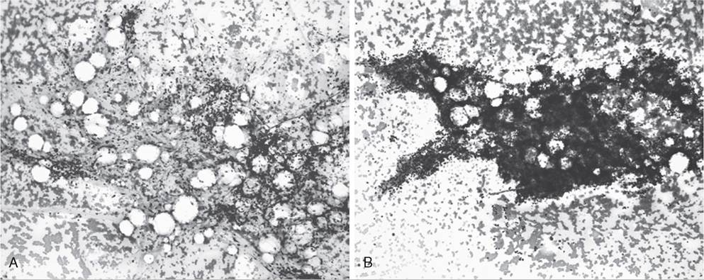
FIG. 28.21 Bone marrow particles. The particle in A is of low cellularity. Only a small proportion of the particle is composed of nucleated cells. The particle in B is of high cellularity. At least 75% of the particle is composed of nucleated cells. (Wright-Giemsa stain, 10? objective.)
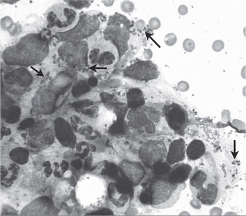
FIG. 28.22 Bone marrow aspirate. The blue-black granules visible extracellularly and within macrophages represent iron stores (arrows). Iron stores can also be seen as brown crystalline material within particles. (Wright-Giemsa stain, 100? objective.)
ratio in a low number of llamas is 0.9 to 2.9.27 Llama, alpaca, and vicuna have a lower M/E ratio at high elevation (4200 m) versus low elevation.2 M/E ratios in horses generally range from 0.5 to slightly greater than 1, with 1.5 sometimes reported as the upper value.10,41-43
Evidence of dysplastic changes in hematopoietic cells includes asynchronous maturation between the cytoplasm and nucleus, large cell forms, and abnormal nuclei, such as ring-shaped nuclei. Cell types other than hematopoietic cells should be evaluated as to their presence and proportion of the total cell population. Low numbers of macrophages (response in the peripheral blood, horses do not. Reticulocytes are rarely seen in equine peripheral blood, and in one investigation it took several days of severe anemia (packed cell volume [PCV] of 13% to 14%) in horses before a reticulocytosis of 1% to 2% was seen.44 An increase in mean corpuscular volume (MCV) may be detected in the peripheral blood of horses with chronic anemia or acute, massive hemolytic or hemorrhagic anemias,43-46 but the best way to assess erythropoiesis in horses is to examine the bone marrow.
The number of reticulocytes significantly increases in the bone marrow of horses with regenerative anemia. In the study of acute blood loss in which only 1% to 2% of reticulocytes were seen in peripheral blood, bone marrow reticulocytes went from 4.1% (pre-blood loss) to as high as 66.5%.44 Reticulocytes typically comprise less than 2% of erythrocytes in normal bone marrow, and a regenerative response is considered present if reticulocytes comprise greater than 5%.15 Determining the number of reticulocytes per 1000 erythrocytes after staining with a supravital stain such as new methylene blue is the most reliable method, but an estimate can be made by counting the number of polychromatophils per 100? oil objective field in a Wright-stained smear (assuming the field of view includes approximately 100 cells; this can vary with the microscope). In the face of anemia, greater than 5 polychromatophils/oil field is considered a good regenerative response, and less than 2 polychromatophils/oil field is indicative of erythropoietic suppression.47 Care should be taken not to dilute the sample with peripheral blood, as this will dilute the number of reticulocytes from the bone marrow.Other bone marrow parameters that indicate erythrocyte regeneration in horses include a decreased M/E ratio (Archer RK: Bone marrow biopsy in the horse: a study of the normal marrow cytology in cross-bred ponies, Vet Rec 66:261, 1954.
17. Murray SL, Lau KW, Begg A, et al: Myelodysplasia, hypophosphataemia, vitamin D, and iron deficiency in an alpaca, Aust Vet J 79:328, 2001.
18. Lawrence WC, Nichols WW, Altera KP: A simple method for bone marrow aspiration in the cow, Cornell Vet 52:297, 1962.
19. Delling U, Lindner K, Ribitsch I, et al: Comparison of bone marrow aspiration at the sternum and the tuber coxae in middle-aged horses, Can J Vet Res 76:52, 2012.
20. Russell KE, Sellon DC, Grindem CB: Bone marrow in horses: indications, sample handling, and complications, Compendium 16:1359, 1994.
21.
Durando MM, Zarucco L, Schaer TP, et al: Pneumopericardium in a horse secondary to sternal bone marrow aspiration, Equine Vet Educ 18: 75, 2006.22. Kisiday JD, Goodrich LR, McIlwraith CW, et al: Effects of equine bone marrow aspirate volume on isolation, proliferation, and differentiation potential of mesenchymal stem cells, Am J Vet Res 74:801, 2013.
23. Kasashima Y, Ueno T, Tomita A, et al: Optimisation of bone marrow aspiration from the equine sternum for the safe recovery of mesenchymal stem cells, Equine Vet J 43:288, 2011.
24. Morris DD: Review of anemia in horses, part I: clinical signs, laboratory findings and diagnosis, Equine Pract 11:27, 1989.
25. Wilde JKH: A technique of bone marrow biopsy in cattle, Res Vet Sci 2:315, 1961.
26. Grunsell CS: Marrow biopsy in sheep, I. normal, Br Vet J 107:16, 1951.
27. Andreasen CB, Gerros TC, Lassen ED: Evaluation of bone marrow cytology and stainable iron content in healthy adult llamas, Vet Clin Pathol 23:38, 1994.
28. Calhoun ML: A cytological study of costal marrow, I. The adult horse, Am J Vet Res 15:181, 1954.
29. Calhoun ML: A cytological study of costal marrow, II. The adult cow, Am J Vet Res 15:395, 1954.
30. Valli VE, Jacobs RM: Structure and function of the hematopoietic system. In Feldman BF, Zinkl JG, Jain NC, editors: Schalm' veterinary hematology, ed 5, Philadelphia, 2000, Lippincott Williams & Wilkins.
31. Wilde JKH: Bovine bone marrow: a note on the total nucleated cell count, Res Vet Sci 4:160, 1963.
32. Ishihara A, Helbig HJ, Sanchez-Hodge RB, et al: Performance of a gravitational marrow separator, multidirectional bone marrow aspiration needle, and repeated bone marrow collections on the production of concentrated bone marrow and separation of mesenchymal stem cells in horses, Am J Vet Res 74:854, 2013.
33. Martin DR, Cox NR, Hathcock TL, et al: Isolation and characterization of multipotential mesenchymal stem cells from feline bone marrow, Exp Hematol 30:879, 2002.
34. Pittenger MF, Mackay AM, Beck SC, et al: Multilineage potential of adult human mesenchymal stem cells, Science 284:143, 1999.
35. Fortier LA, Potter HG, Rickey EJ, et al: Concentrated bone marrow aspirate improves full-thickness cartilage repair compared with microfracture in the equine model, J Bone Joint Surg Am 92:1927, 2010.
36. Burton AG, Clark KC, Borjesson DL, et al: Equine bone marrow volume reduction, red blood cell depletion, and mononuclear cell recovery using the PrepaCyte-CB processing system, Vet Clin Pathol 44:188, 2015.
37. Bourzac C, Smith LC, Vincent P, et al: Isolation of equine bone marrow- derived mesenchymal stem cells: a comparison between three protocols, Equine Vet J 42:519, 2010.
38. Schalm AO, Lasmanis J: Cytologic features of bone marrow in normal and mastitic cows, Am J Vet Res 37:359, 1976.
39. Wilde JKH: The cellular elements of the bovine bone marrow, Res Vet Sci 5:213, 1964.
40. Winqvist G: Morphology of the blood and the hematopoietic organs in cattle under normal and some experimental conditions. The bovine bone marrow, Acta Anat (Basel) 22(Suppl 21):33, 1954.
41. Jain NC: Examination of the blood and bone marrow, Philadelphia, 1993, Essentials of veterinary hematology Lea & Febiger.
42. Latimer KS, Andreasen CB: Bone marrow. In Cowell RL, Tyler RD, editors: Cytology and hematology of the horse, Goleta, California, 1992, American Veterinary Publications.
43. Schalm OW 1975. Bone marrow erythroid cytology in anemias of the horse. Proceedings of the First International Symposium on Equine Hematology American Association of Equine Practitioners: Golden.
44. Tablin F, Weiss L: Equine bone marrow: a quantitative analysis of erythroid maturation, Anat Rec 213:202, 1985.
45. Lumsden JH, Valli VE, McSherry BJ, et al: The kinetics of hematopoiesis in the light horse, II. The hematological response to hemorrhagic anemia, Can J Comp Med 39:324, 1975.
46. Radin MJ, Eubank MC, Weiser MG: Electronic measurement of erythrocyte volume and volume heterogeneity in horses during erythrocyte regeneration associated with experimental anemias, Vet Pathol 23:656, 1986.
47. Schalm OW: Equine hematology: part IV erythroid marrow cytology in response to anemia, Equine Pract 2:35, 1980.
48. Franken P, Wensing T, Schotman AJH: The bone marrow of the horse, II. Warm-blooded horses with anaemia, Zentralbl Veterinarmed A 29:23, 1982.
49. Malikides N, Kessell A, Hodgson JL, et al: Bone marrow response to large volume blood collection in the horse, Res Vet Sci 67:285, 1999.
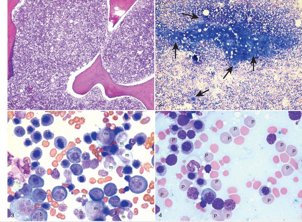
Color Plates 28.1 to 28.4 Bone marrow biopsy and aspirate from a 5-year-old female Quarter Horse with marked anemia (packed cell volume [PCV] = 9%; 31%-47%) and thrombocytopenia (42,000 platelets/gL; 125,000-300,000/gL). The bone marrow is highly cellular as seen in Color Plates 28.1 (H&E) and 28.2 (Wright-Geimsa); arrows point to megakaryocytes, 10? objectives. The myeloid/erythroid (M/E) ratio is decreased at 0.3, and polychromatophils comprise 30% of red blood cells (RBCs). Color Plate 28.3 shows a large erythroid population (unmarked cells and b = erythroblast) with fewer myeloid cells (m) and lymphocytes (L); many polychromatophils (P) can be seen in Color Plate 28.4 (Wright-Geimsa, 100? objective). This is representative of regeneration and consistent with either chronic blood loss or immune-mediated destruction of erythrocytes, which could both be occurring in this horse. The horse was diagnosed with intramuscular hemangiosarcoma.
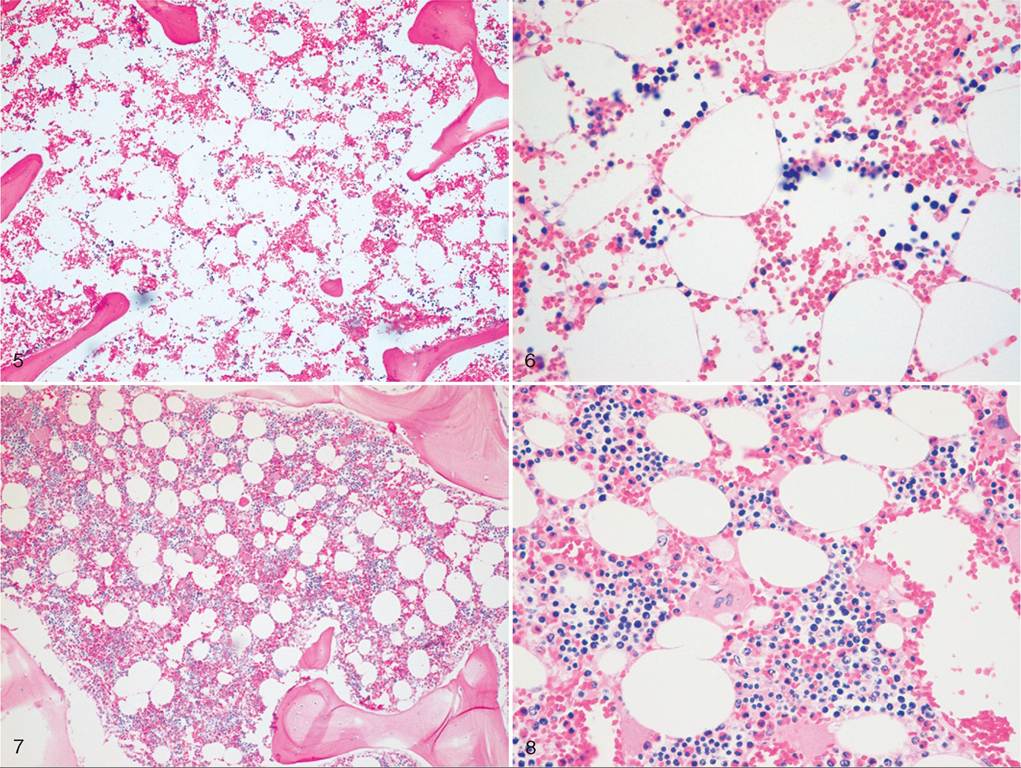
Color Plates 28.5 to 28.8 Bone marrow biopsy from a 2-year-old mixed-breed steer with mild anemia (packed cell volume [PCV] = 23%; 24%-46%), severe neutropenia (0/gL; 600-4000/gL), and thrombocytopenia (3000 platelets/gL; 100,000-800,000/gL) (Color Plates 28.5 and 28.6), compared to a normal age-matched control (Color Plates 28.7 and 28.8). Severe generalized bone marrow hypoplasia is apparent. The marked bone marrow suppression in this case is due to bracken fern toxicity. H&E; 10? (Color Plates 28.5 and 28.7) and 40? (Color Plates 28.6 and 28.8) objectives.
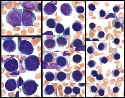
Color Plate 28.9 Maturational stages of erythrocytes within the bone marrow. The left panel contains rubriblasts in the upper square with prorubricytes below. Rubriblasts are large cells with large nuclei containing coarse-grained chromatin and nucleoli; a perinuclear halo is present within scant, deeply basophilic cytoplasm. Prorubricytes have a more mature nucleus; chromatin condensation begins, and no nucleoli are present. The middle panel contains basophilic and polychromatophilic rubricytes; chromatin condensation is distinct, and the cytoplasm is paler, grayer, or polychromatic. The right panel contains metarubricytes with dark, intense chromatin condensation and pink cytoplasm. (Wright-Giemsa stain, 100? objective.)
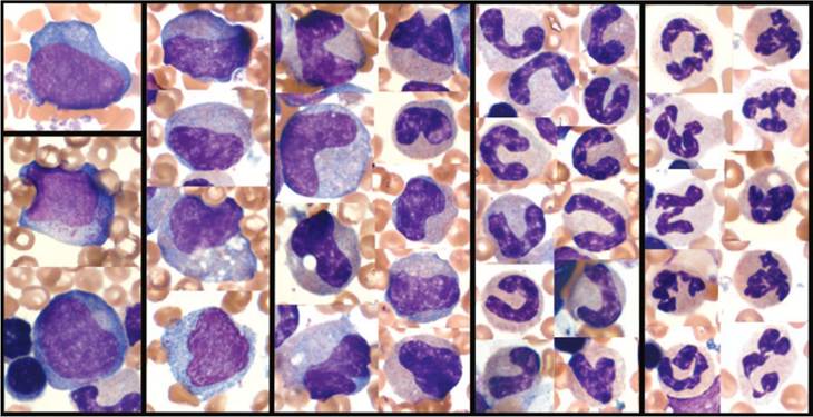
Color Plate 28.10 Maturational stages of granulocytes within the bone marrow. The left panel contains a myeloblast in the upper square with promyelocytes below. Myeloblasts are large cells with large nuclei containing fine reticular chromatin and nucleoli; the cytoplasm is basophilic and agranular. Promyelocytes contain coarse cytoplasmic granulation; nuclear chromatin is beginning to condense, and nucleoli are present. Subsequent panels, moving to the right, contain myelocytes, metamyelocytes, band neutrophils, and segmented neutrophils. Myelocytes have a lower nuclear/cytoplasmic ratio and contain fewer and finer cytoplasmic granules; the nucleus is round to kidney-shaped and contains no nucleoli. As maturation progresses, the cells become smaller, the cytoplasm becomes more eosinophilic, the chromatin becomes more condensed, and the nucleus indents and segments.