Causative Agents of Adiaspiromycosis and Relatives
Adiaspiromycosis is a pulmonary infection by members of the family Ajellomycetaceae (order Onygenales) where the fungus is present in pulmonary tissue as very large, thick-walled resting cells known as adiaspores (Fig.
7.1). The genus Emmonsia was described for fungi producing adiaspores and until recently comprised two species: Emmonsia parva and E. crescens (Ciferri and Montemartini 1959; Emmons and Jellison 1960). Emmonsia pasteuriana was later added to the genus (Drouhet et al. 1998) despite the absence of adiaspores, instead having a pathogenic phase with principally small budding cells. Recent molecular phylogenetic studies have recognised the polyphyletic nature of the genus Emmonsia, and consequent revisions have changed the taxonomic landscape considerably (Dukik et al. 2017; Jiang et al. 2018). Classical mating experiments demonstrated that E. crescens has an Ajellomyces teleomorph (Sigler 1996), and DNA sequence comparisons already had suggested a sister species relationship between Emmonsia parva (the type species of Emmonsia) and Blastomyces dermatitidis (Peterson and Sigler 1998; Sigler 2005). With the validation of the name Blastomyces (de Hoog et al. 2017), the generic name Emmonsia has been discarded as a synonym. Emmonsia parva was thus reclassified as a Blastomyces species, B. parvus (Jiang et al. 2018). Emmonsia crescens was described as the type species of a separate genus, Emmonsia (Jiang et al. 2018). Emmonsia crescens is responsible for most animal cases of adiaspiromycosis (Sigler 2005), a disease which is occasionally observed in humans (Anstead et al. 2012). Emmonsia pasteuriana was also reclassified as the type species of a new genus, Emergomyces (E. pasteurianus), alongside Emergomyces africanus (Kenyon et al. 2014; Dukik et al. 2017), Emergomyces orientalis (Wang et al. 2017), Emergomyces canadensis and Emergomyces europaeus (Jiang et al. 2018). Emergomyces pasteurianus and E. africanus, which both have a yeast rather than an adiaspore tissue form, principally cause disseminated infections in humans with T-cell immune defects (Drouhet et al. 1998; Feng et al. 2015; Schwartz et al. 2015a; Malik et al. 2016; Dukik et al. 2017).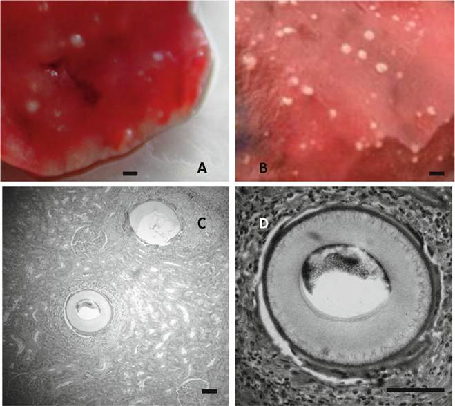
Fig. 7.1 Adiaspiromycosis in mammalian lungs. Gross lesions of adiaspiromycosis in the lung of a mole (a), and an otter (b). (c, d) Histopathological section of experimentally infected monkey kidney (inoculation directly into kidney) showing adiaspores surrounded by granulomata. H&E stain. Scale bar = 1 mm (Panels A and B) or 100 μm (Panels C and D)
7.2
More on the topic Causative Agents of Adiaspiromycosis and Relatives:
-
Veterinarian -